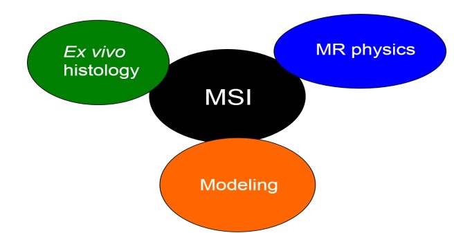
Figure 1: Micro Scale Imaging or MSI consists of three complementary pillars. MSI combines cutting-edge techniques in MR physics (supported by advance image processing [3.4]) with mathematical models of the MR signal [1.2,5] to measure histological biomarkers in vivo. For validation, MSI directly compares the measured in vivo biomarkers with gold standard markers from ex vivo histology.
Emmy Noether Project: "Validating MRI-based in-vivo histology"
This project will develop a novel in-vivo magnetic resonance imaging (MRI) framework for measuring white-matter micro-scale compartments in the human brain, which were previously only accessible by ex-vivo histology. Micro Scale Imaging or MSI (see also Fig. 1) will work at an unprecedented spatial resolution of 500 microns, i.e. at the volume level a factor of 64 higher than standard qMRI data (~2 mm). It will measure white-matter properties, including myelin density, axonal density, and the g-ratio [1]. These properties are of direct functional relevance because they are related to the conduction speed of nerve fibers and thus important for the communication between anatomical areas.
To this end, MSI will use biophysical models to relate the signal of five different quantitative MRI techniques (relaxometry, diffusion MRI, magnetization transfer, quantitative phase, and proton density imaging) to the proposed micro-scale metrics [2]. The models make use of the fact that the signal of each of these techniques is differently influenced by the micro-scale compartments and thus offers complementary information (e.g., some quantitative MRI techniques are more sensitive to axonal orientation while others are more sensitive to myelin or fiber densities).
To find the best biophysical metric for the g-ratio, myelin density, and fiber density, my group will quantitatively compare the respective MSI metric and other established biophysical metrics with the corresponding marker from ex-vivo histology.
Vacancies: One Postdoc and one PhD positions
References
[1] Mohammadi S, Carey D, Dick F, Diedrichsen J, Sereno MI, Reisert M, Callaghan MF and Weiskopf N (2015), Frontiers in Brain Imaging Methods, Whole-brain in-vivo measurements of the axonal g-ratio in a group of 37 healthy volunteers , 9: 00441 .
[2] Weiskop Nf, Mohammadi S, Lutti A, Callaghan MF (2015) Advances in MRI-based computational neuroanatomy: from morphometry to in-vivo histology , Curr Opin Neurol. 28(4):313-22 .
[3] Tabelow K (*), Mohammadi S (*), Weiskopf N, Polzehl J (2015) POAS4SPM: a toolbox for SPM to denoise diffusion MRI data. Neuroinformatics 13:1929 . (*) Shared first authorship.
[4] Ruthotto L, Mohammadi S, Weiskopf N (2014) A new method for joint susceptibility artefact correction and super-resolution for dMRI. Med Imaging Imaging Proc 9034:90340 P-90340 P-4 .
[5] Callaghan M, Pine K, Tabelow K, Polzeh J, Weiskopf N, Mohammadi S (2016)
Mapping Higher Order Components of the GRE Signal Decay at 7T with Short TE Data through Adaptive Smoothing In Proc Intl Soc Magn Reson Med. 2016;24:1539.