Bioengineering / Medical Technology
Bioengineering / Bone Quality / Tissue and Material Analytics
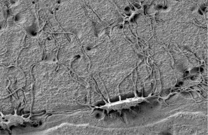
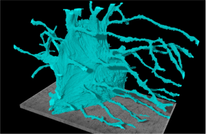
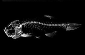
The fracture risk of bone is not solely dependent on bone quantity; indeed, bone’s hierarchical structure has distinct features at multiple length-scales, and thus the quality of the structure plays a large role in its resistance to fracture. Understanding how the nature of fracture is affected by bone quality requires the characterization of various compositional and morphological features at each level of bone’s complex hierarchical structure, which has characteristic features at multiple length-scales, specifically evolving to its macroscopic form (>3 mm) from a nanostructure comprised of collagen and mineral (<500 nm) and a microstructure of lamellae, osteocyte lacunae (3 to 20 µm) and osteons (100 to 300 µm). We believe that such a characterization of bone will lead to a greater understanding of how aging and disease affect bone’s structural features and increase fracture risk. The unique feature of our studies is the analysis of the ultrastructure in physiologic and pathologic conditions. We use an integrated approach combining bone quality assessment and medical technology techniques by using microcomputed tomography (Micro-CT), bone histomorphometry, synchrotron small-angle X-ray scattering (SAXS) / wide-angle X-ray diffraction (WAXD), backscattered electron imaging (BSE), microanalysis (EDX, µXRF), Nanoindentation, Raman/Fourier Transform Infrared spectroscopy (FTIR) and high performance liquid chromatography (HPLC) in order to characterize various levels of the hierarchical structure of skeletally intact and diseased bones, whose mechanical properties are determined via materials testing. These measurements can be carried out with imaging of how the crack paths specifically interact with the bone-matrix components (SEM), thereby providing insight into the controlling toughening and damage mechanisms involved. In this connection, we investigate whether the ultrastructure in terms of the nature of the collagen and mineral characteristics is significantly altered in bone diseases, whether there are distinct changes in structural and material characteristics, and how these changes can affect the mechanisms of fracture. Our aim is to provide new information on how distinct ultrastructural features affect the mechanical behavior of bone tissue; additionally, as this is a departure from purely medical studies of bone fracture, we hope that such bioengineering and materials-science-based studies can positively impact the medical field by providing new and different insights into bone-related diseases, and as such can help in the search for new cures and treatment options for bone diseases.
Principal Investigator

Univ.-Prof. Dr. rer. medic. Björn Busse
Head of Division, Bioengineering / Medical Technology
Multiscale Imaging / Analytics / Bone and Tissue Quality
Group Members


Praveer Sihota, Ph.D.
Post-Doc
Multiscale analysis of bone composition and material properties