Precision Made in Rome
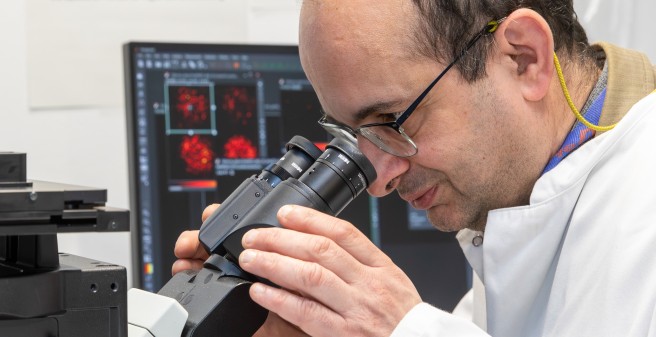
Dr. Antonio Virgilio Failla has been leading the UKE Microscopy Imaging Facility (UMIF) since 2011, where he and his team develop novel imaging technologies and train researchers to use them. Before coming to Hamburg, the Rome-born physicist worked at research institutions in Heidelberg, Potsdam, Tübingen and Cambridge.
Text: Nicole Sénégas-Wulf, Photos: Eva Hecht
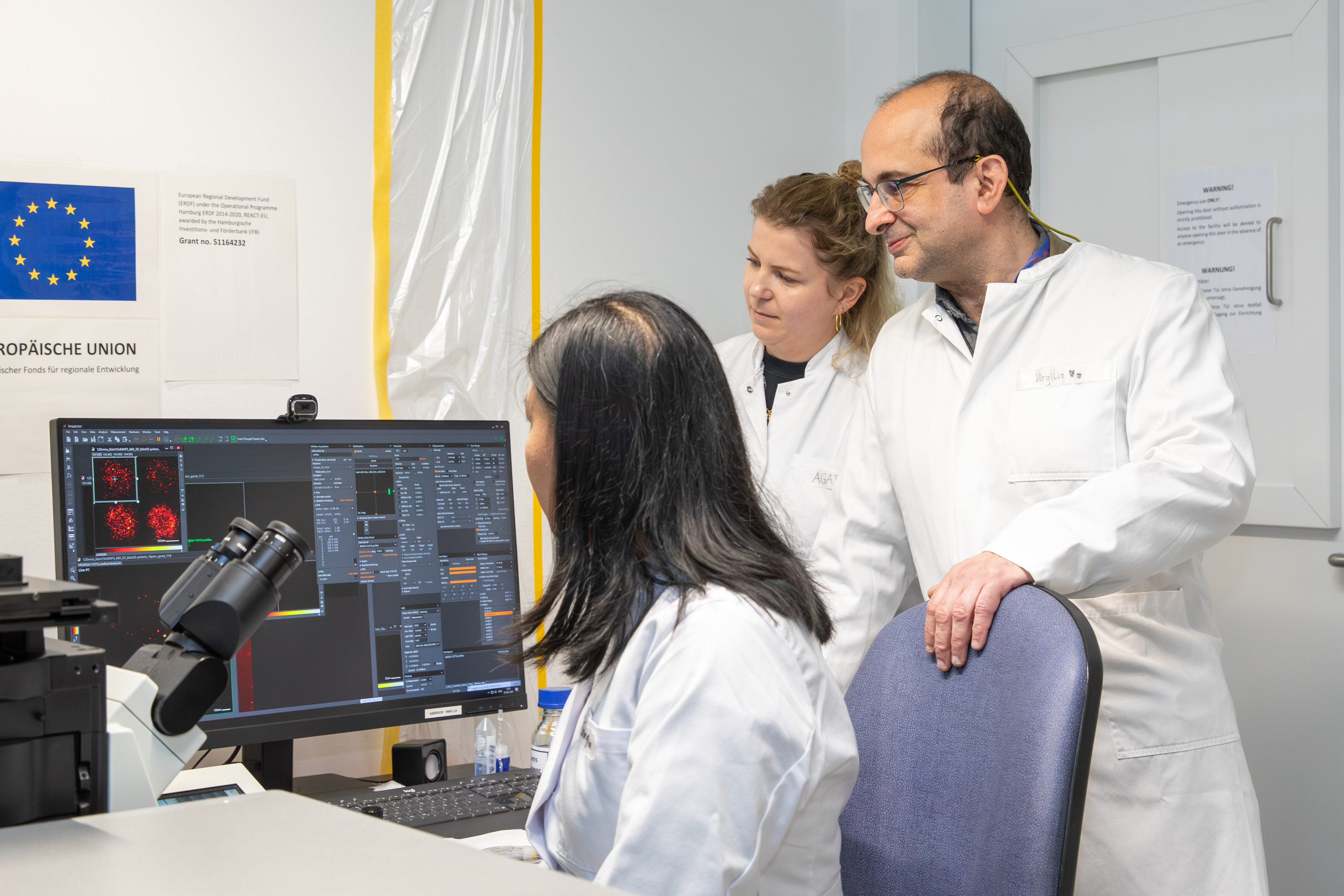
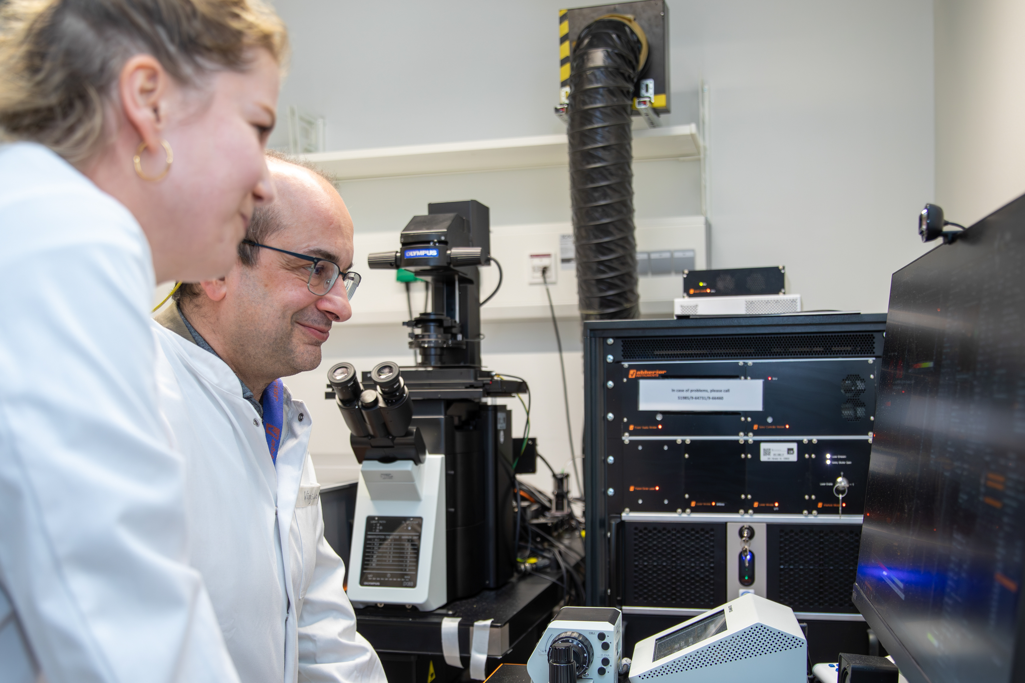
In the UMIF office on the ground floor of Campus Research I, a small basketball hoop hangs to the right of the door, just below the ceiling. What’s it for? Dr. Failla laughs: “When researchers register to use one of our microscopes, they first have to test their shooting skills and make the shot.” A bit of humor helps foster a sense of community, says Dr. Failla with a smile, adding that he sees himself as a service provider to the scientific community. “I don’t do biomedical research myself, but I develop methods that help UKE researchers drive their work forward.” His area of expertise is bioimaging – the visualization of biological structures using super-resolution microscopes. Altogether, he and his team of three manage 13 systems at Campus Research.
The language of biology
Bioimaging has always fascinated him. After studying physics in Rome with a focus on optics, he moved to Heidelberg, where he developed a wide-field microscope as part of his doctoral research between 2002 and 2003, making tiny cellular structures visible. After working in Potsdam and Tübingen, he spent two years in Cambridge at a cancer research UK imaging lab. “That’s where I learned to work with biological samples – and to speak the language of biologists,” Dr. Failla says. This has been invaluable in his role at the UKE. “I didn’t study biology or medicine, but I understand exactly what biologists want when they come to me.” After returning to Germany, he became facility manager in Tübingen before moving to the UKE in Hamburg in 2011, where he has since developed the microscopy unit into a leading center for advanced imaging.
In the service of science
“Part of my work involves developing new technologies,” says the 54-year-old. Recently, he and his team published a new method for diagnosing Alzheimer’s disease as a less invasive alternative. The UMIF is also active in coronavirus research and is currently working to visualize how the virus interacts with other cells using different staining techniques. More recently, Dr. Failla has focused on the original application of super-resolution light microscopy on biomedical samples. New developments include in-house image analysis algorithms for novel bioimaging experiments, as well as algorithms based on artificial intelligence and deep learning. In addition to research, training is a key part of Dr. Failla’s role. “In our sessions, we teach UKE staff the fundamentals of microscopy and offer courses to help researchers master specialized techniques.” Confident, simple use of the technology is essential for producing accurate, meaningful results, he says.

Between saber and saw
Why has he spent so many years in Germany? “Firstly, because it’s a top destination for microscopy. Secondly, because I simply like Germany.” Over the years, he has built many meaningful relationships here – for example with his former football team in Potsdam, which he still keeps in touch with. “That’s also where I learned German – on the pitch,” he says with a wink. And, of course, through his wife, who is German. While he no longer plays football actively, he continues to practice saber fencing, in which he has even become a veteran German team champion. Outside of sports, Dr. Failla can often be found in his home workshop, where he enjoys designing and building his own furniture – from bookshelves to beds. His most recent project: an incubator for his garden plants. A bit of biology is always a must.