Project - A05:
T cells and cholangiocytes: an interaction that governs portal immune regulation and determines biliary inflammation
Project leader:
Dr. rer. nat. Dorothee Schwinge

Summary
Autoimmune cholangiopathies are progressive disorders of bile ducts leading to cholestasis with fibrosis and subsequent biliary cirrhosis. Lack of pathogenetic understanding and limited therapeutic options make cholangiopathies a major indication for liver transplantation. Thus, effective and novel treatment options are urgently needed. The etiology of cholangiopathies is largely unknown, but several mechanisms have been considered to be part of a complex and multifactorial disease pathogenesis, including dysregulated adaptive immune responses, autoimmunity and defects in mechanisms protecting against bile acid toxicity. A central mechanism in the pathogenesis of cholangiopathies is an immune response directed against cholangiocytes leading to cholangiocyte damage, proliferation and neo-ductuli formation. There is emerging evidence that cholangiocytes are not only targets, but also actively modulate immune cell function via cell-to-cell contact or soluble mediators. Thus, activated cholangiocytes participate in inflammation by secreting chemokines and cytokines that are involved in recruitment and crosstalk with immune, vascular and mesenchymal cells. However, little is known about the antigen-presenting function of cholangiocytes. We could recently show that T cell-induced upregulation of PD-L1 on cholangiocytes limits experimental cholangitis severity. Within this project we now aim to decipher the impact of cholangiocytes on T cell responses and to investigate the ability of cholangiocytes to convey antigen-presenting functions. Moreover, we will determine the consequences of T cell-cholangiocyte interaction for the activation, regeneration and senescence of cholangiocytes. Overall, we hypothesise that the bidirectional interaction between T cells and cholangiocytes determines biliary inflammation and thus the pathogenesis of cholangiopathies. We expect these experiments to delineate the functional role of cholangiocytes in shaping the portal immune regulation and to identify novel treatment targets for autoimmune cholestatic liver diseases.
Project plan
Immune responses directed against cholangiocytes are central to the pathogenesis of immune mediated and autoimmune cholangiopathies. Portal infiltrates in the livers of people with PBC and PSC are predominantly composed of T cells. We therefore believe that T cells significantly contribute to ductal inflammation, leading to periductal fibrosis and progression of the disease. There is emerging evidence from our and other groups that cholangiocytes are not only inactive targets, but also actively promote disease progression by secreting cytokines and chemokines as well as expression of co-stimulatory and co-inhibitory molecules. In this project, we hypothesise that the bidirectional interaction between T cells and cholangiocytes determines biliary inflammation and is central to portal immune regulation.
In order to test this hypothesis, our work programme has the following work packages (WP):
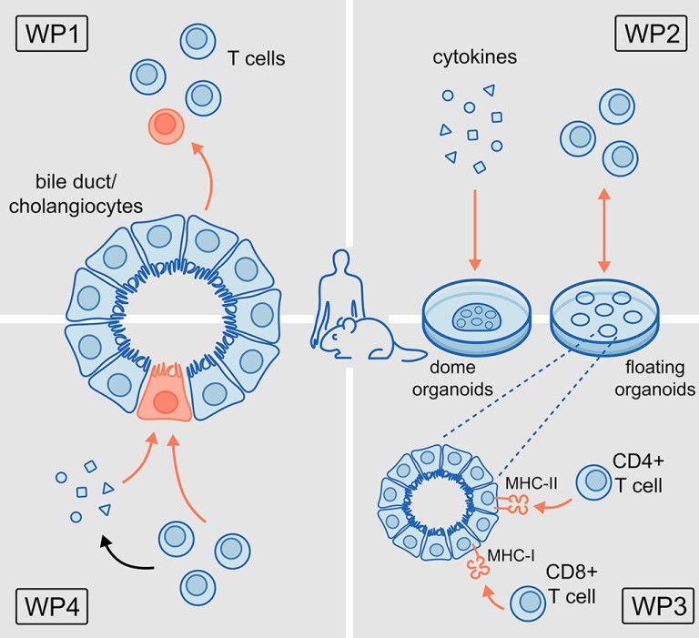
WP1: To identify the modulatory capacity of cholangiocytes on T cell proliferation, plasticity and differentiation in vivo using well-established mouse models of acute cholangitis.
WP2: To investigate the bidirectional network between T cells and cholangiocytes in in vitro co-culture models using murine and human cholangiocyte organoids.
WP3: To decipher the antigen presenting capacity of cholangiocytes.
WP4: To characterise the impact of T cells and cytokines on the activation, regeneration and senescence of cholangiocytes using established in vivo models of chronic cholangitis and in vitro cell culture systems.
Project related publications
Katt J, Schwinge D, Schoknecht T, Quaas A, Sobottka I, Burandt E, Becker C, Neurath MF, Lohse AW, Herkel J, Schramm C. Increased T helper type 17 response to pathogen stimulation in patients with primary sclerosing cholangitis. Hepatology 2013;58:1084-1093. doi: 10.1002/hep.26447. Epub 2013 Jul 30.
Kunzmann LK#, Schoknecht T#, Poch T, Henze L, Stein S, Kriz M, Grewe I, Preti M, Hartl J, Pannicke N, Peiseler M, Sebode M, Zenouzi R, Horvatits T, Böttcher M, Petersen BS, Weiler-Normann C, Hess LU, Ahrenstorf AE, Lunemann S, Martrus G, Fischer L, Li J, Carambia A, Kluwe J, Huber S, Lohse AW, Franke A, Herkel J, Schramm C, Schwinge D. Monocytes as Potential Mediators of Pathogen-Induced T-Helper 17 Differentiation in Patients With Primary Sclerosing Cholangitis (PSC). Hepatology 2020;72:1310-1326. doi: 10.1002/hep.31140. Open access.
Poch T#, Krause J#, Casar C, Liwinski T, Glau L, Kaufmann M, Ahrenstorf AE, Hess LU, Ziegler AE, Martrus G, Lunemann S, Sebode M, Li J, Schwinge D, Krebs CF, Franke A, Friese MA, Oldhafer KJ, Fischer L, Altfeld M, Lohse AW, Huber S, Tolosa E#, Gagliani N#, Schramm C#. Single-cell atlas of hepatic T cells reveals expansion of liver-resident naive-like CD4+ T cells in primary sclerosing cholangitis. J Hepatol 2021; 24: S0168-8278(21)00219-1. doi:10.1016/j.jhep.2021.03.016. Open access.
Schmidt T, Schwinge D, Rolvien T, Jeschke A, Schmidt C, Neven M, Butscheidt S, Kriz M, Kunzmann L, Mussawy H, Hubert J, Hawellek T, Rüther W, Oheim R, Barvencik F, Lohse AW, Schramm C, Schinke T, Amling M. Th17 cell frequency is associated with low bone mass in primary sclerosing cholangitis. J Hepatol 2019;70:941-953. doi:10.1016/j.jhep.2018.12.035. Open access.
Schoknecht T, Schwinge D, Stein S, Weiler-Normann C, Sebode M, Mucha S, Otto B, Ellinghaus E, Stahl F, Franke A, Lohse AW, Herkel J, Schramm C. CD4+ T cells from patients with primary sclerosing cholangitis exhibit reduced apoptosis and down-regulation of proapoptotic Bim in peripheral blood. J Leukoc Biol 2017;101:589-597.doi: 10.1189/jlb.5A1015-469R. Epub 2016 Sep 14. Open access.
Sebode M#, Peiseler M#, Franke B, Schwinge D, Schoknecht T, Wortmann F, Quaas A, Petersen BS, Ellinghaus E, Baron U, Olek S, Wiegard C, Weiler-Normann C, Lohse AW, Herkel J, Schramm C. et al. Reduced FOXP3(+) regulatory T cells in patients with primary sclerosing cholangitis are associated with IL2RA gene polymorphisms. J Hepatol 2014;60:1010-1016. doi:10.1016/ j.jhep.2013.12.027. Epub 2014 Jan 8. Open access.
Schwinge D#, von Haxthausen F#, Quaas A, Carambia A, Otto B, Glaser F, Höh B, Thiele N, Schoknecht T, Huber S, Steffens N, Lohse AW, Herkel J, Schramm C. Dysfunction of hepatic regulatory T cells in experimental sclerosing cholangitis is related with IL-12 signalling. J Hepatol 2017;66:798-805. doi:10.1016/j.jhep.2016.12.001. Open access.
Glaser F#, John C#, Engel B#, Höh B#, Weidemann S, Dieckhoff J, Stein S, Becker N, Casar C, Schuran FA, Wieschendorf B, Preti M, Jessen F, Franke A, Carambia A, Lohse AW, Ittrich H, Herkel J, Heeren J, Schramm C#, Schwinge D#. Liver infiltrating T cells regulate bile acid metabolism in experimental cholangitis. J Hepatol 2019;pii:S0168-8278(19)30347-2. doi: 10.1016/j jhep. 2019.05.030. Open access.
Schwinge D, Carambia A, Quaas A, Krech T, Wegscheid C, Tiegs G, Prinz I, Lohse AW, Herkel J, Schramm C. Testosterone Suppresses Hepatic Inflammation by the Downregulation of IL-17, CXCL-9, and CXCL-10 in a Mouse Model of Experimental Acute Cholangitis. J Immunol 2015;194:2522-30. doi: 10.4049/jimmunol.1400076 . Epub 2015 Feb 11.
Stein S, Henze L, Poch T, Carambia A, Krech T, Preti M, Schuran FA, Reich M, Keitel V, Fiorotto R, Strazzabosco M, Fischer L, Li J, Müller LM, Wagner J, Gagliani N, Herkel J, Schwinge D#, Schramm C#. IL-17A/F enable cholangiocytes to restrict T cell-driven experimental cholangitis by upregulating PD-L1 expression. J Hepatol 2020:S0168-8278(20)33759-4. doi: 0.1016/j.jhep.2020. 10.035. Open access.
#equally contributing authors