Project - B04:
ILC development and modulation of cholangiocytes in biliary atresia
Project leader:
Prof. Dr. Madeleine J. Bunders, MD/PhD
Summary
The immune system undergoes dramatic changes after birth. The invasion of trillions of microbes requires adequate host-defense responses. At the same time, organs that suddenly face a high microbial load, such as the liver, need to generate sufficient tolerogenic responses to prevent severe tissue inflammation. In addition to these challenges our group and others have furthermore shown that immune cells importantly contribute to tissue development early in life. In some children the complex demands on the immune system early in life are not met, resulting in severe inflammation and sclerosis of the biliary tract and liver, a disease called biliary atresia (BA). Due to the lack of effective treatment, the majority of children with BA progress to severe liver failure. BA is the number one indication for paediatric liver transplantation worldwide. We have previously shown that tissue-resident innate lymphocytes (ILCs) are critical mediators of tissue development and adaption to the extra-uterine environment in the intestine. However, our understanding of the development of ILCs in the liver of children during this important phase in life is limited due to the lack of paediatric studies. To address this gap, we performed preliminary analyses of paediatric and adult liver-derived ILCs, including children with BA and children requiring a liver transplant due to a diverse range of underlying metabolic diseases (non-inflamed paediatric livers). These data suggest that ILCs are enriched in paediatric compared to adult livers. Furthermore, in the livers of children with BA, frequencies of pro-inflammatory IL-17-producing ILCs were increased compared to non-inflamed livers of children without BA. Pro-inflammatory ILCs are known to contribute to immune-mediated diseases and damage epithelial barriers. To investigate the effects of ILCs on cholangiocytes in BA, we generated 3D cholangiocyte organoids and observed that IL-17 stimulation of cholangiocytes from children with BA negatively affected their proliferative capacity while upregulating phenotypic markers associated with fibrosis. Based on these preliminary findings, we hypothesise that ILC development is dysregulated in BA and contributes to cholangiocyte pathology in young children. Building on our expertise in organ-specific human immune development and modelling of ILC-epithelial cell interactions in 3D organoids, we will investigate ILC development in the liver after birth and how ILC dysregulation impacts cholangiocytes in the development and progression of BA. The results of this project will reveal age-specific ILC development in human livers and identify targets for therapeutic approaches to restore tissue homeostasis in BA-affected livers.
Project plan
Our central hypothesis is that ILCs play an important role in homeostasis of the liver after birth and are dysregulated in BA, where they further enhance cholangiocyte dysregulation. Based on this central hypothesis, we aim to investigate the development of ILCs in the liver and identify age-specific ILCs that contribute to tissue homeostasis after birth, but are dysregulated in the context of BA. Building on our studies showing that ILCs are critical regulators in intestinal tissue development, we will study the role of ILCs in liver development and their contribution to cholangiocyte dysregulation in BA. The long-term aim is to restore homeostatic ILC-cholangiocyte interactions and promote tissue healing of the bile ducts by inhibiting ILC-mediated tissue damage.
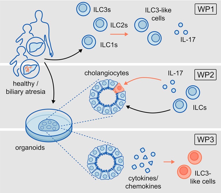
These studies will be performed in the following work packages (WP):
WP1: To characterise ILC development in the liver in children.
WP2: To investigate the role of ILCs in regeneration of cholangiocytes during physiological human liver development in comparison to fibrosis in BA.
WP3: To identify the pathways in BA-affected cholangiocytes that alter the microenvironment of ILCs and promote conversion to ILC3-like cells in BA.
Project related publications
Ziegler AE, Fittje P, Müller LM, Ahrenstorf AE, Hagemann K, Hagen SH, Hess LU, Niehrs A, Poch T, Ravichandran G, Löbl SM, Padoan B, Brias S, Hennesen J, Richard M, Richert L, Peine S, Oldhafer KJ, Fischer L, Schramm C, Martrus G, Bunders MJ, Altfeld M, Lunemann S. The co-inhibitory receptor TIGIT regulates NK cell function and is upregulated in human intrahepatic CD56bright NK cells. Front Immunol 2023;14:1117320. doi: 10.3389/fimmu.2023.1117320. Open access.
Hess LU, Martrus G, Ziegler AE, Langeneckert AE, Salzberger W, Goebels H, Sagebiel AF, Hagen SH, Poch T, Ravichandran G, Koch M, Schramm C, Oldhafer KJ, Fischer L, Tiegs G, Richert L, Bunders MJ, Lunemann S, Altfeld M. The Transcription Factor Promyelocytic Leukemia Zinc Finger Protein Is Associated With Expression of Liver-Homing Receptors on Human Blood CD56bright Natural Killer Cells. Hepatol Commun 2020;4:409-424. doi: 10.1002/hep4.1463. Open access.
Lunemann S, Martrus G, Goebels H, Kautz T, Langeneckert A, Salzberger W, Koch M, Bunders MJ, Nashan B, Van Gisbergen KPJM, Altfeld M. Hobit expression by a subset of human liver-resident CD56bright Natural Killer. Sci Rep 2017;7:6676. doi: 10.1038/s41598-017-06011-7. Open access.
Zecher BF, Ellinghaus D, Schloer S, Niehrs A, Padoan B, Baumdick ME, Yuki Y, Martin MP, Glow D, Schröder-Schwarz J, Niersch J, Brias S, Müller LM, Habermann R, Kretschmer P, Früh T, Dänekas J, Wehmeyer MH, Poch T, Sebode M, Ellinghaus E, Degenhardt F, Körner C, Hoelzemer A, Fehse B, Oldhafer KJ, Schumacher U, Sauter G, Carrington M, Franke A, Bunders MJ, Schramm C, Altfeld M. HLA-DPA1∗02:01∼B1∗01:01 is a risk haplotype for primary sclerosing cholangitis mediating activation of NKp44+ NK cells. Gut 2024;73:325-337. doi: 10.1136/gutjnl-2023-329524. Open access.
Sagebiel AF, Steinert F, Lunemann S, Körner C, Schreurs RRCE, Altfeld M, Perez D, Reinshagen K, Bunders MJ. Tissue-resident Eomes + NK cells are the major innate lymphoid cell population in human infant intestine. Nat Commun 2019;10:975. doi: 10.1038/s41467-018-08267-7. Open access.
Möller KJ*, Wegner LHM*, Malsy J*, Baumdick ME, Borggrewe M, Jordan-Paiz A, Jung JM, Martrus G, Kretschmer P, Sagebiel AF, Schreurs RRCE, Hagen SH, Burmester G, Clauditz TS, Pals ST, Boettcher M, Melling N, Sauter G, Tomuschat C, Königs I, Schumacher U, Altfeld M, Bernink JH, Perez D, Reinshagen K, Bunders MJ. Expanded ILC2s in human infant intestines promote tissue growth. Mucosal Immunol 2023;16:408-421. doi: 10.1016/j.mucimm.2023.04.004. Open access.
Schreurs RRCE, Baumdick ME, Sagebiel AF, Kaufmann M, Mokry M, Klarenbeek PL, Schaltenberg N, Steinert FL, van Rijn JM, Drewniak A, The S-MML, Bakx R, Derikx JPM, de Vries N, Corpeleijn WE, Pals ST, Gagliani N, Friese MA, Middendorp S, Nieuwenhuis EES, Reinshagen K, Geijtenbeek TBH, van Goudoever JB, Bunders MJ. Human fetal TNF-α-cytokine-producing CD4 + effector memory T cells promote intestinal development and mediate inflammation early in life. Immunity 2019;50:462-476.e8. doi: 10.1016/j.immuni.2018.12.010. Open access.
Baumdick ME, Niehrs A, Degenhardt F, Schwerk M, Hinrichs O, Jordan-Paiz A, Padoan B, Wegner LHM, Schloer S, Zecher BF, Malsy J, Joshi VR, Illig C, Schröder-Schwarz J, Möller KJ; Hamburg Intestinal Tissue Study Group; Martin MP, Yuki Y, Ozawa M, Sauter J, Schmidt AH, Perez D, Giannou AD, Carrington M, Davis RS, Schumacher U, Sauter G, Huber S, Puelles VG, Melling N, Franke A; International Inflammatory Bowel Disease Genetics Consortium; Altfeld M*, Bunders MJ*. HLA-DP on Epithelial Cells Enables Tissue Damage by NKp44+ Natural Killer Cells in Ulcerative Colitis. Gastroenterology 2023;165:946-962.e13. doi: 10.1053/j.gastro.2023.06.034. Open access.
Bunders MJ, Altfeld M. Implications of Sex Differences in Immunity for SARS-CoV-2 Pathogenesis and Design of Therapeutic Interventions. Immunity 2020;53:487-495. doi: 10.1016/j.immuni.2020. 08.003. Open access.
# equally contributing authors