Research at the UKE?
 DE
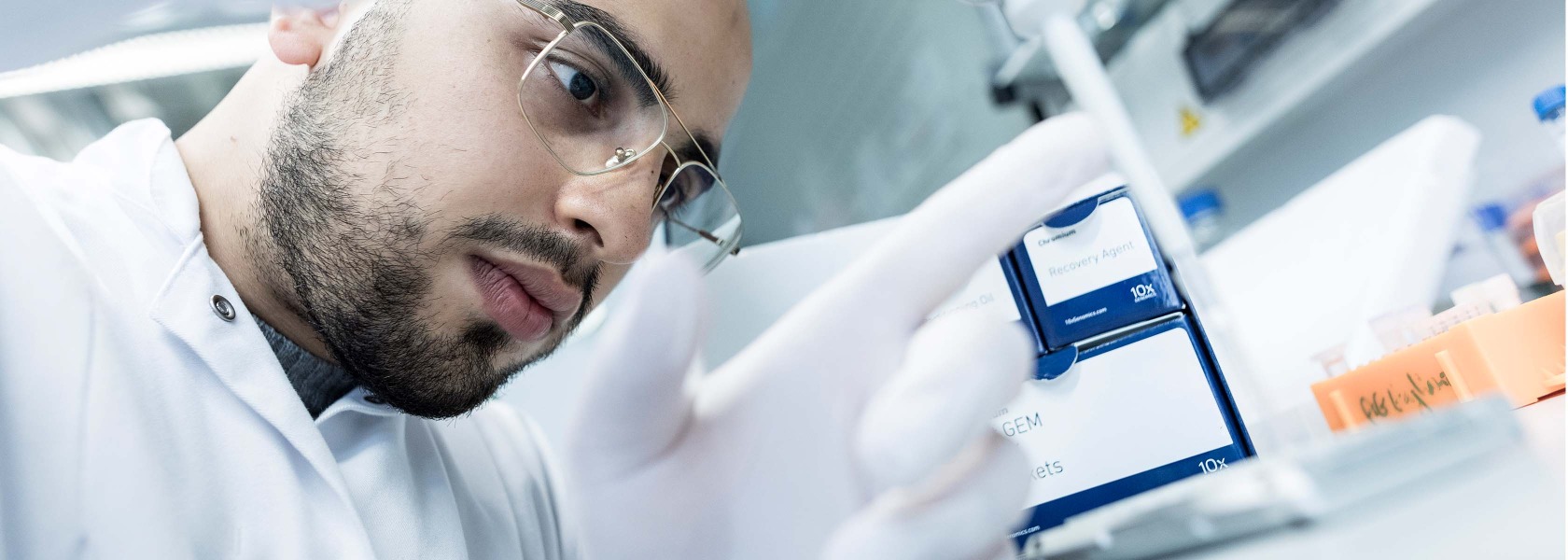

Institute for Inflammation and Carcinogenesis
We like studying the basic molecular and cellular mechanisms of T cell biology, adaptation to environmental changes, and ultimately, how T cells
either mediate physiological responses to pathogens or pathological tissue damaging processes.

“Unraveling the complexities of CD4+ T cell biology in immune-related disorders.”
Prof. Dr. Nicola Gagliani

“Harnessing the power of the immune system to fight cancer.”
Dr. Joseph Tintelnot
Project details and goals
Our laboratory focuses on unraveling the complexities of CD4 T cell biology, in particular their metabolism, adaptability, and roles in disease.
We investigate the different CD4 T cell subsets by exploring their developmental trajectories and the molecular mechanisms underlying their adaptability.
A key aspect of our research is understanding how CD4 T cells adapt to environmental changes, including both classical immune challenges like infections and non-classical stimuli such as dietary shifts.
We are particularly interested in the organ-specific responses of resident CD4 T cells and their implications for immune adaptation. Finally, we study the involvement of CD4 T cells in immune-mediated inflammatory diseases and cancer, profiling their interactions with other immune cells, for example macrophages, and with the different tissues at a single-cell level.
Through these efforts, we aim to contribute to a deeper understanding of CD4 T cell function and their potential in treating immune-related disorders.
Current projects
Basic Aspects of CD4+ T Cell Biology
CD4+ T cells are a plastic and heterogeneous population made up of subsets that differ in cytokine production, cell surface markers, function and tissue specificity. Although there have been efforts to classify these subsets based on surface markers and signature cytokines, unsupervised analysis of CD4+ T cell populations shows an unexpected heterogeneity which is not reflected by the classical textbook nomenclature. Supporting this, the use of cytokine fate-mapping mouse models revealed that some CD4+ T cell subsets can adapt their functionality during the development of the immune response. Redefining the classification of CD4+ T cells by understanding their plastic developmental trajectories and identifying the epigenetic and molecular mechanisms controlling these trajectories are some of the main topics of our group.
Immune adaptation of CD4+ T cells to environmental changes
CD4+ T cells possess the ability to switch from a pro- to an anti-inflammatory function, i.e. immune adaptation. The consequences of dysfunctional immune adaptation are clear: the immune cells either fail to mount a protective response to lethal infections, or they overreact to non-harmful antigens and, in doing so, induce a variety of immune-mediated inflammatory diseases (Xu H. et al Nat Comm 2020).
CD4+ T cells adapt in response to both “classical” immune stimuli, such as infections, and “non-classical” immune stimuli, such as changes in dietary habits. In addition, the immune adaptation of CD4+ T cells varies across different organs. There is indeed a conspicuous number of resident CD4+ T cells that have established an intimate and diverse relationship with each organ, which is not yet fully understood.
Our lab is particularly interested in decoding the organ-specific response of resident CD4+ T cells to different classical and non-classical stimuli, with a particular focus on the diet.

The role of CD4+ T cells in immune-mediated inflammatory diseases and cancer
CD4+ T cells are key players in orchestrating a context-appropriate immune response, but their protection comes at a price. They can, in fact, also promote immune-mediated inflammatory diseases (IMIDs), including the development of inflammation-induced carcinogenesis. By profiling resident T cells and their interactions with other immune and non-immune cells in a broad spectrum of diseased human organs at single-cell resolution, we are delineating a new anthology of T cell function in IMIDs and cancer. Using in vitro 3D culture, we validate the newly discovered functions of these cells and provide the scientific and clinical community with new targets for potential future immune therapies.
Publications 2026
Liu B, Spalinger MR, Invernizzi A, Gerdes E, Steglich B, Schwarzfischer M, Machicote A, Pelczar P, Pöhlmann D, Nawrocki M, Böttcher M, Aleko A, Velasquez LN, Wende S, Stallbaum F, Neuendorff J, Grosshauser S, Muscate F, Douilhet G, Perez LG, Sabihi M, Möller K. Nat Commun. 2026 Apr 18. doi: 10.1038/s41467-026-71808-y. Online ahead of print.PMID: 42009642
Abstract
Tofacitinib, a pan-Janus kinase inhibitor, and the Janus kinase 1-preferential inhibitors Upadacitinib and Filgotinib are approved for the treatment of ulcerative colitis, yet their molecular mechanisms of action remain incompletely understood. Here, using dextran sulfate sodium-induced and T cell transfer colitis models together with analyses of individuals with ulcerative colitis, we show that all three inhibitors ameliorate colitis in mice with macrophage-specific deletion of protein tyrosine phosphatase non-receptor type 2, a model characterized by hyperactive Janus kinase-signal transducer and activator of transcription signaling. In contrast, only Upadacitinib and Filgotinib provide enhanced protection in wild-type mice - an effect that is lost upon genetic disruption of inflammasome signaling. Longitudinal single-cell transcriptomic analyses and immunostaining of intestinal biopsies further show that Upadacitinib reduces interleukin-1β expression in vivo, which associates with clinical response. Thus, indirect suppression of inflammasome activity contributes to the efficacy of Janus kinase 1-preferential inhibitors.
Siracusa F, Gagliani N. Immunity. 2026 Mar 10;59(3):512-514. doi: 10.1016/j.immuni.2026.02.010
Abstract
Immune responses are energetically costly and must adapt to systemic nutrient availability. How immunity is reorganized to preserve host glucose during caloric restriction remains unclear. In this issue of Immunity, Menezes-Silva et al. show that dietary restriction, via glucocorticoids, rewires innate and adaptive immunity, and this leads to the conservation of glucose while maintaining protective host defense.
Xu Y, Weltzsch JP, Kilian C, Steglich B, Weiler-Normann C, Dudek M, Fackler J, Wehmeyer MH, Tintelnot J, Liebig LA, Steinmann S, Laschtowitz A, Horst LJ, Schregel I, Sebode M, Hartl J, Casar C, Lu J, Schön G, Zapf A, Bono MR, Rosemblatt MV, Nuñez S, Castañeda J, Weidemann SA, Kaiser N, Schwerk M, Kolster M, Rattay G, Ulrich H, Sivayoganathan V, Song N, Krause J, Böttcher M, Sagebiel A, Wagner J, Krebs CF, Puelles VG, Hübner N, Tolosa E, Bonn S, Huber S, Knolle PA, Herkel J, Adlung L, Schramm C, Gagliani N, Lohse AW. J Hepatol. 2026 Mar 19:S0168-8278(26)00120-0. doi: 10.1016/j.jhep.2026.02.026. Online ahead of print.PMID: 41864242
Abstract
Background & aims: Patients with autoimmune hepatitis (AIH) experience increased mortality and severe side effects from non-specific immunosuppressive therapy, highlighting an urgent need for targeted treatment approaches. Here, we aimed to delineate the cellular and molecular network underlying AIH within its spatial context and to validate a key therapeutic target in a clinical trial.
Methods: We employed computational modelling, multi-omics analyses, and functional experiments to map the immune landscape of AIH. In addition, we conducted a steroid-free open-label phase IIa clinical trial using infliximab, a TNF-targeting antibody, in patients with AIH.
Results: Our studies revealed that myeloid cell and hepatocyte-derived IL-15 promotes cytotoxicity and proliferation of liver auto-aggressive CD8+ T cells. Full execution of their cytotoxic program is licensed by TNF derived from clonally expanded liver-resident CD4+ T cells. AIH hepatocytes respond to TNF by increasing expression of adhesion molecules, making them targets for both CD8+ and CD4+ T cells. In the clinical trial, targeting TNF with infliximab demonstrated efficacy as an entirely steroid-free AIH treatment.
Conclusions: These findings elucidate the immune network in AIH and identify TNF as one of the central network nodes. Accordingly, our findings provide the basis for novel targeted, steroid-free immune therapies, including the use of infliximab.
Clinical trial number: European Union Clinical Trials Register (EudraCT No.: 2017-003311-19).
Impact and implications: These findings have significant implications for the treatment of autoimmune hepatitis (AIH). By mapping the spatial and functional immune network within the AIH liver, this study identifies IL-15 and TNF as central drivers of T cell-mediated cytotoxicity, offering new precision targets for intervention. The successful use of infliximab as a steroid-free therapy in a phase II trial marks a pivotal step toward safer, more specific treatment options for patients with AIH. This research not only advances our understanding of AIH pathogenesis, but also sets the stage for broader application of immune-targeted therapies in autoimmune liver diseases.
Siracusa F, Machicote A, Huber S, Gagliani N. Curr Opin Immunol. 2026 Feb;98:102713. doi: 10.1016/j.coi.2025.102713. Epub 2025 Dec 22
Abstract
The intestinal microbiota transforms dietary components into bioactive metabolites that profoundly influence mucosal and systemic immunity. Short-chain fatty acids, secondary bile acids and tryptophan-derived indoles are among the most studied microbial metabolites shaping T, B and innate immune cell functions through a variety of mechanisms, including receptor signaling, epigenetic modification and metabolic reprogramming. Dietary habits strongly affect the composition of the intestinal microbiota and thus, the production and availability of these microbial metabolites, with consequences that range from protective immune regulation to detrimental inflammatory responses. Here, we review recent findings from mouse and human studies, highlighting how the microbiota–immunity axis can be modulated by diets and discuss implications for tissue homeostasis, infection and chronic inflammatory diseases. Understanding this complex interplay may guide the development of ad hoc dietary and microbial interventions to restore tolerance and improve therapeutic outcomes.
Siracusa F, Gagliani N. Trends Immunol. 2026 Jan;47(1):1-2. doi: 10.1016/j.it.2025.11.003. Epub 2025 Nov 19
Abstract
Chi and colleagues revealed that dietary cysteine enhances intestinal stem cell (ISC) regeneration, driving coenzyme A (CoA) synthesis and expansion of intraepithelial (IEL) CD8αβ+ T cells that secrete IL-22. This epithelial-immune crosstalk potentiates ISC repair after injury, highlighting a metabolism-immune axis linking cysteine sensing to tissue regeneration.
Publications 2025
Thiele K, Urbschat C, Riquelme JIA, Ahrendt LS, Wöhrle R, Schepanski S, Eckert JJ, Becht E, Qi M, Alawi M, Becker M, Gagliani N, Mittrücker HW, Diemert A, Arck PC. Nat Commun. 2025 Jul 15;16(1):6522. doi: 10.1038/s41467-025-61572-w.PMID: 40664658
Abstract
Subsequent pregnancies are generally less prone to obstetric complications. A successful pregnancy outcome requires pivotal immunological adaptation to ensure immune tolerance towards the foetus. Thus, the lower risk for pregnancy complication during subsequent pregnancies may be attributable to immune memory mounted during first pregnancies. Here we identify higher frequencies of fetal-antigen-specific CD4+ regulatory T (Treg) cells both postpartum and in subsequent pregnancies in mice which are partly originating from trans-differentiated Th17 cells. Our functional experiments demonstrate that these CD4+ Treg cells have memory functions (CD4+ mTreg) and account for an improved fetal development and pregnancy outcome, also during adverse conditions, such as gestational sound stress. Using a high-throughput single-cell quantification method, we identify candidate markers for the detection of CD4+ mTreg cells, which include CXCR4 and CD274. Our findings thus contribute to the improved understanding of pregnancy-induced immune memory and foster the identification of immune targets aiming to reduce the risk for immune-mediated pregnancy complications.
Asada N, Ginsberg P, Paust HJ, Song N, Riedel JH, Turner JE, Peters A, Kaffke A, Engesser J, Wang H, Zhao Y, Khatri R, Gild P, Dahlem R, Diercks BP, Das S, Ignatova Z, Huber TB, Prinz I, Gagliani N, Mittrücker HW, Krebs CF, Panzer U. Nat Immunol. 2025 Apr;26(4):557-566. doi: 10.1038/s41590-025-02105-x. Epub 2025 Mar 6.PMID: 40050432
Abstract
Tissue-resident memory T (TRM) cells are a specialized T cell population that reside in tissues and provide a rapid protective response upon activation. Here, we showed that human and mouse CD4+ TRM cells existed in a poised state and stored messenger RNAs encoding proinflammatory cytokines without protein production. At steady state, cytokine mRNA translation in TRM cells was suppressed by the integrated stress response (ISR) pathway. Upon activation, the central ISR regulator, eIF2α, was dephosphorylated and stored cytokine mRNA was translated for immediate cytokine production. Genetic or pharmacological activation of the ISR-eIF2α pathway reduced cytokine production and ameliorated autoimmune kidney disease in mice. Consistent with these results, the ISR pathway in CD4+ TRM cells was downregulated in patients with immune-mediated diseases of the kidney and the intestine compared to healthy controls. Our results indicated that stored cytokine mRNA and translational regulation in CD4+ TRM cells facilitate rapid cytokine production during local immune response.
Song Z, Klyuchnikov E, Badbaran A, Tan L, Dress RJ, Czajkowski E, Weßler S, Massoud R, Wolschke C, Schimrock A, Zhang Y, Ly C, Gagelmann N, Rathje K, Fehse B, Bonn S, Ravens S, Gagliani N, Krebs CF, Panzer U, Ayuk F, Kröger N, Prinz I. Biomark Res. 2025 Dec 1;13(1):155. doi: 10.1186/s40364-025-00868-x.
Abstract
Background: Alloreactive T cells mediate graft-versus-leukemia (GvL) reactions and acute graft-versus-host disease (aGvHD) in AML patients following allogeneic hematopoietic stem cell transplantation.
Methods: To investigate biomarkers that identify alloreactive T cells associated with either beneficial GvL or detrimental aGvHD, we collected graft samples and two post-transplant follow-up blood samples (day 30 and day 100) of ten AML patients undergoing hematopoietic stem cell transplantation and profiled over 777,000 CD45+ leukocytes in total by combinatorial barcoding-based mega-scale single-cell RNA sequencing.
Results: Using immune receptor sequences as intrinsic clonal barcodes, we observed that especially CD8+ graft-derived T cells persisted and displayed enhanced proliferation, clonal expansion, and likely alloreactivity. Notably, patient-derived peripheral leukocytes that survived the conditioning, as identified by sex-chromosome-related genes, were primarily CD4+ T helper cells. MDGA1 expression on T cells and NK cells emerged as a novel biomarker potentially associated with aGvHD. Additionally, we observed a significant deficiency of ADGRG1 expression, a marker of alloreactive cytotoxic T cells, by αβ and γδ T cells from relapsed patients.
Conclusions: In conclusion, mega-scale single-cell monitoring of graft and hematopoietic immune cell reconstitution allowed us to demonstrate that MDGA1 and ADGRG1 may function as complementary biomarkers expressed by distinct circulating T cells that are associated with divergent outcomes in AML patients, enabling precise risk stratification of alloHSCT outcomes and presenting potential therapeutic targets.
Publications
Explore more of our recent and previous publications
Team


Prof. Dr. med. Nicola Gagliani
Head
E-mail address: