
Liver Lab – Immune regulation
Liver inflammation is a physiological response to injury that is necessary for the healing and regeneration of the organ. However, a dysregulated inflammatory response can lead to liver disease. Our research projects aim to better understand the regulation of inflammation in the liver in order to therapeutically target mechanisms involved.
Particular focus is on autoimmune responses, which can be helpful to remove damaged or premalignant liver cells, but can also cause autoimmune liver diseases, when dysregulated.

"The physiological mechanisms of the liver can be harnessed to induce antigen-specific immune tolerance for the treatment of autoimmune diseases."
Principal investigator

"Identifying the mechanisms that lead to the accumulation of disease specific autoantibodies will gain novel insights in the pathogenesis of autoimmune liver diseases."

“The liver is unique in many ways – one of them is its complex immunoregulatory role. Under physiological conditions the liver favours tolerance, but at the same time it can be the site of every immune-mediated diseases. To understand the rules that govern this spectrum need to be understood to develop more targeted immunotherapies, and to maintain health.”
Prof. Dr. med. Ansgar W. Lohse
"The microbiota is an important determinant of our health. We investigate how intestinal and biliary microbiota influence the regulation of immunity in the liver and thus will identify novel therapeutic targets."
Prof. Dr. med. Christoph Schramm

"Understanding how autoimmunity is regulated in the liver will pave the way towards new therapies for liver diseases."
Prof. Dr. rer. nat. Johannes Herkel

"Deciphering the functional role of cholangiocytes in shaping portal immune regulation will help identify novel treatment targets for autoimmune cholestatic liver diseases."
Dr. rer. nat. Dorothee Schwinge
Project details and goals
Immune cells continually survey the liver to check the organ’s state of health, and protect it from infections or malignancy. Aberrations from a healthy state can cause liver inflammation causing the removal of aberrant cells, followed by restoration of a healthy liver state. However, the regulation of liver inflammation and its resolution can go wrong, causing the development of chronic inflammation and autoimmune liver diseases.
There are three autoimmune liver diseases, autoimmune hepatitis, primary sclerosing cholangitis and primary biliary cholangitis, causing life-long inflammation of the liver parenchyma or bile ducts. All three diseases have an unmet need for more specific, effective and tolerable treatments.
Yet in healthy state, the liver can also down-regulate inflammatory activities of immune cells, which can be harnessed for antigen-specific induction of immune tolerance and the treatment of autoimmune diseases affecting other organs.
Our research aims to elucidate the immune regulation in the liver, both in autoimmune liver diseases and in liver tolerance. We believe that understanding hepatic immune regulation is key to the development of novel therapies for autoimmune diseases of the liver and other organs. Indeed, our strategy has led to the development of two novel therapeutic approaches currently being tested in clinical trials.
Current projects
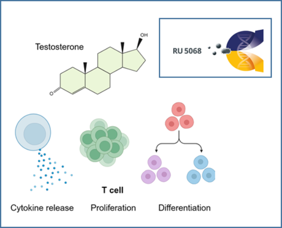
The role of androgens in autoimmune liver diseases
The autoimmune liver diseases (AILD) autoimmune hepatitis (AIH) and primary biliary cholangitis (PBC) exhibit some of the highest female predominance among all autoimmune diseases. Despite this notable sex imbalance, the underlying reasons for it remain unknown. There is mounting evidence that androgens play an important role in mediating sex differences in immunity. To investigate the effects of androgens on immune cells, we analysed cohorts of transgender people receiving gender-affirming hormone therapy (GAHT) and age- and sex-matched people with PBC, AIH and healthy controls. Our results show that testosterone has direct effects on T cell function and improves disease course in a patient with AILD. We believe that the elucidation of the role of androgens in the context of AILD will improve our understanding of autoimmune liver diseases.
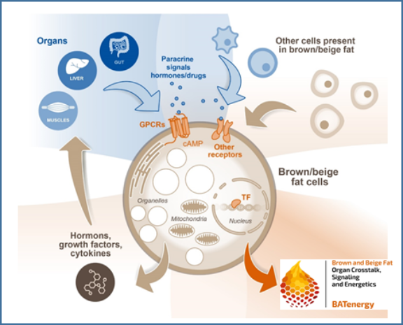
Intestinal metabolites and their impact on thermogenic responses by brown and white adipose tissues
Microbial metabolites such as short chain fatty acids (SCFA) and secondary bile acids are assumed to play a major role in the regulation of energy metabolism of the host. Recently, we discovered a novel immunometabolic crosstalk between activated T cells in the liver and hepatocytes that decreased the expression of enzymes regulating bile acid metabolism. Here, transfer of antigen-specific T cells resulted in increased bile acids serum levels, which potentially could trigger energy expenditure. Within this project we hypothesize that hepatocellular handling of gut-derived metabolites and the immunometabolic interaction between hepatocytes and T cells in the liver control energy uptake and expenditure by thermogenic brown and beige adipocytes.
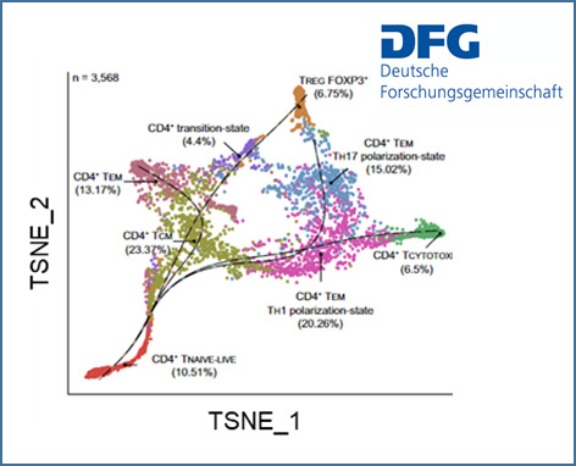
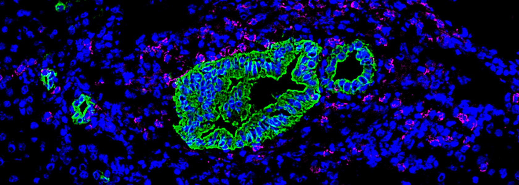
Composition and functional heterogeneity of T cells in primary sclerosing cholangitis (PSC)
Primary sclerosing cholangitis (PSC) is an enigmatic liver disease characterised by a chronic inflammation leading to scarring and obliteration of intra- and extrahepatic bile ducts. The pathogenesis is largely unknown but it is widely believed that PSC is an immune mediated disease with environmental and genetic factors contributing to immune dysregulation. We recently identified an intrahepatic naive-like population of CD4+ T cells in people with PSC that is prone to polarize towards a pro-inflammatory phenotype. Moreover, we found that a gene polymorphism in the BACH2 gene significantly impacts CD4+ T cell differentiation.
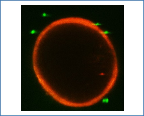
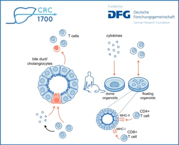
Immune cells and cholangiocytes: an interaction that determines biliary inflammation
Autoimmune cholangiopathies such as primary sclerosing cholangitis (PSC) and primary biliary cholangitis (PBC) are progressive disorders of bile ducts leading to cholestasis with fibrosis and subsequent biliary cirrhosis. A central pathogenetic mechanism is the immune response directed against cholangiocytes, the epithelial cells lining the bile ducts. Chronic inflammation in cholangiopathies leads to cholangiocyte damage, proliferation and neo-ductuli formation. Within this project we hypothesize that the bidirectional interaction between immune cells and cholangiocytes determines biliary inflammation. We expect these experiments to delineate the functional role of cholangiocytes in shaping the hepatic immune cell function and to identify novel treatment targets for autoimmune cholestatic liver diseases.
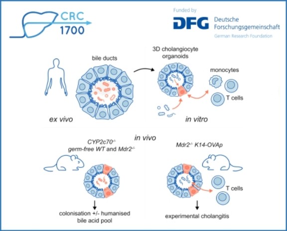
The role of intestinal and biliary microbiota in autoimmune liver diseases
Primary sclerosing cholangitis (PSC) is a disease characterised by immune dysregulation at mucosal surfaces and thus ideally suited to study the role of biliary mucosal barrier function for hepatic immune regulation. It has been shown that PSC is characterised by mucosal and (peri-)ductular inflammation of bile ducts that results in periductular fibrosis and ultimately biliary cirrhosis. Cholangiocytes are the lining cells of bile ducts that form the biliary mucosal barrier. They modify the composition of bile fluid, but also transport information from the biliary endoluminal environment to the portal tracts of the liver, including intrahepatic resident and migrating immune cell populations. Importantly, the biliary tree represents a large mucosal surface and clinical observations suggest that recurrent bacterial cholangitis and potentially also colonisation of the bile ducts with Candida sp. aggravates PSC disease course. Within this project we aim to understand how biliary microbiota affect cholangiocyte function and the interaction of cholangiocytes with immune cells. This understanding will enable novel therapeutic strategies aimed at restoring homeostasis at the mucosal biliary barrier.
In addition, we study the role of intestinal microbiota in autoimmune liver diseases and how they influence inflammatory activity and symptom burden of disease.
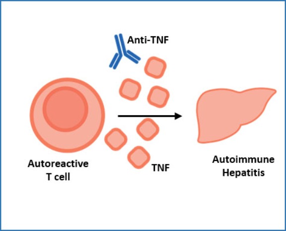
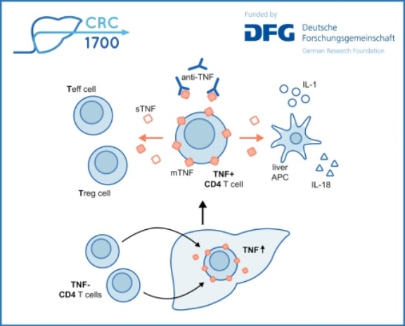
The role of TNF in autoimmune hepatitis
CD4+ T cells in the livers of autoimmune hepatitis (AIH) patients produce high levels of the cytokine TNF, and AIH can be treated with anti-TNF antibodies. What exactly TNF does in AIH livers is unclear, as TNF can have different effects in different contexts. Here we aim to elucidate the disease-causing mechanisms of TNF in AIH, and to identify additional treatment targets
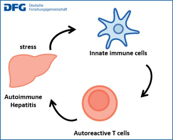
Dysregulation of autoimmunity in autoimmune hepatitis: mechanisms and experimental therapies
Autoimmune hepatitis (AIH) is presumably caused by an immune response to liver antigens. In healthy people, such immunity to autoantigens is regulated by diverse mechanisms to prevent sustained organ damage. Here, we analyse the regulation of autoimmunity in AIH patients to identify dysregulated mechanisms that might be corrected therapeutically. To that end, we characterise the state of and interactions between liver cells, innate immune cells and lymphocytes. The aim is to find better therapies for AIH.
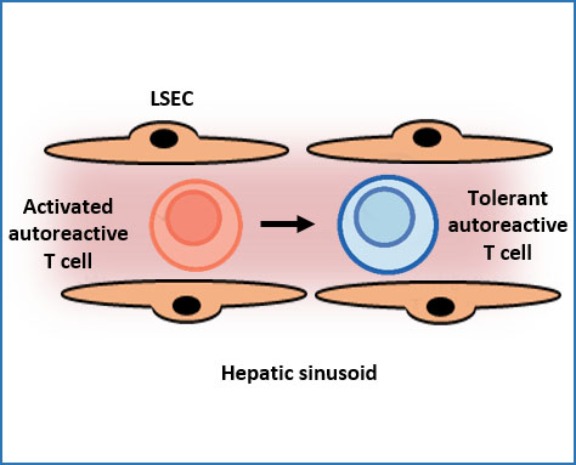
Immune tolerance induction in the hepatic sinusoids
The immune system attacks most antigens and substances that are deemed harmful to the body. The liver, however, has a remarkable ability to suppress inflammation and damaging immune activities directed to antigens in the liver. This so called hepatic tolerance can prevent inflammatory diseases, but also promote chronic infections or cancer. Here, we aim to elucidate the mechanisms of hepatic tolerance in the hepatic sinusoids, in order to develop new therapies by manipulating hepatic tolerance.
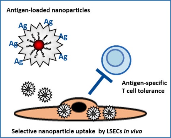
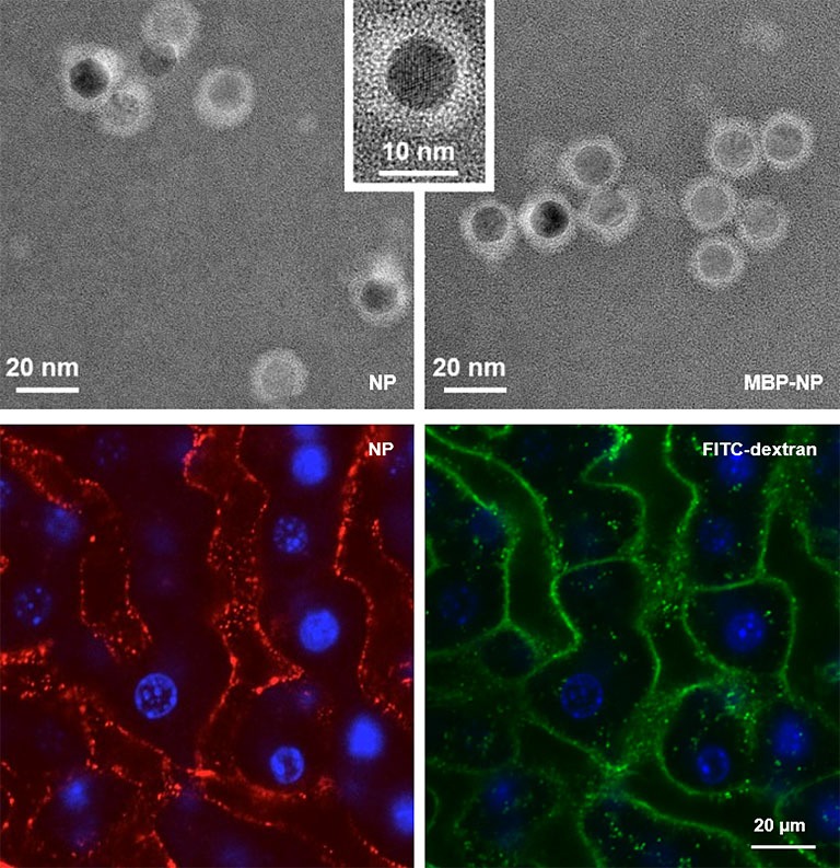
A nanoparticle-based therapy platform for antigen-specific tolerance induction in autoimmune diseases
Endothelial cells in the hepatic sinusoids (LSECs) are major inducers of hepatic tolerance. LSECs are scavenger cells that can take up blood-borne antigens with high capacity and present those to T cells, which then become tolerant to these antigens. We developed a nanoparticle-based therapy platform targeting LSECs for antigen-specific tolerance induction. This approach was successfully applied in various preclinical inflammation models and is currently tested in clinical trials. We will further study the applicability of this promising approach.
Publications 2026
Al Jawazneh A, Liebold I, Leyk S, Lanzloth C, Brackrock V, López-López T, Graute S, Aranda-Pardos I, Hamley M, Bahn J, Heeren J, Von Felden J, Lohse AW, Schramm C, Schwinge D, Sáez PJ, A-Gonzalez N, Huber S, Jacobs T, Adlung L, Worthmann A, Bosurgi L. J Exp Med. 2026 Jul 6;223(7):e20242079. doi: 10.1084/jem.20242079. Epub 2026 Jun 23. PMID: 42334420; PMCID: PMC13289589.
Abstract
The clearance of apoptotic cells by phagocytes is crucial for restoring tissue balance after injury. In autoimmune liver diseases like primary sclerosing cholangitis, cell death is thought to result from accumulation of toxic bile acids within parenchymal cells. Whether, in this context, bile acid–loaded dying cells impact the efficiency of phagocytic macrophages in restoring tissue balance remains unknown. Here, we demonstrate that in a murine model of cholangitis, bile acids accumulate in a subpopulation of efferocytic macrophages with pro-inflammatory features. Our in vitro results indicate that, upon their engulfment, apoptotic hepatocytes laden with bile acids can serve as Trojan horses, delivering bile acids into efferocytic macrophages and thereby shaping macrophage function. This contrasts with the characteristics of macrophages that engulf apoptotic parenchymal cells lacking bile acids. Together, our findings delineate a system in which the content of the phagocytosed dying cells, specifically bile acid–laden hepatocytes, drives a pro-inflammatory program in the corresponding efferocytic macrophages, potentially contributing to chronic hepatic inflammation.
Gottwick C, Averhoff P, Casar C, Liebig LA, Pilz SM, Haas V, Krzikalla D, Fleischer S, Hübner N, Adlung L, Schwinge D, Schramm C, Carambia A, Herkel J. Front Immunol. 2026 May 5;17:1834595. doi: 10.3389/fimmu.2026.1834595. PMID: 42164495; PMCID: PMC13183527.
Abstract
Background and aims: Liver sinusoidal endothelial cells (LSECs) have a key role in maintaining organismal homeostasis by scavenging blood-borne molecules and inducing specific immune tolerance to ingested antigens. The scavenger and tolerance function of LSECs can be harnessed for specific treatment of autoimmune diseases by nanoparticle-mediated autoantigen delivery to LSECs. In liver fibrosis, which is a frequent condition in human populations, LSECs undergo changes promoting pro-fibrotic and pro-inflammatory activation of other hepatic cells, but it is unclear whether the scavenger and immune tolerance functions of LSECs are affected.
Methods: Utilizing two mouse models of liver fibrosis, we explored the ability of LSECs to take up nanoparticles conjugated with antigen peptides, to present the ingested antigen peptides to T cells and to induce peptide-specific immune tolerance in vitro and in vivo in the context of autoimmune diseases.
Results: LSECs from fibrotic livers showed few distinct adaptations regarding immune functions; however, overall LSEC identity was largely maintained. Accordingly, endocytosis of nanoparticles by LSECs in vivo, as well as processing and presentation of nanoparticle-bound antigen peptides was not compromised by liver fibrosis. LSECs from fibrotic livers maintained the ability to effectively induce the generation of regulatory T cells from conventional CD4 T cells. Hence, targeted delivery of autoantigen peptides to LSECs in vivo effectively induced specific tolerance despite liver fibrosis, providing protection in two models of experimental autoimmune disease. Analysis of datasets from human subjects with or without liver cirrhosis confirmed that scavenger and tolerance pathways in LSECs were preserved in human liver fibrosis.
Conclusions: Scavenger activity and antigen-specific tolerance induction by LSECs are preserved in liver fibrosis. Thus, LSECs remain reliable mediators of homeostasis and tolerance under fibrotic conditions, and particularly suitable targets for nanomedicine products.
Evangelakos I, Verkade E, Rohde JK, Zaufel A, Vargek M, Heine M, Worthmann A, Graute S, Manka Fuh M, Gunasekaran K, Kumari M, Schwinge D, von Bergen M, Rolle-Kampczyk U, Engelmann B, Breinbauer R, Fuerst R, Saleem U, de Boer JF, Schlein C, Hansen A, Scheja L, Kuipers F, Moustafa T, Heeren J. Mol Metab. 2026 May;107:102363. doi: 10.1016/j.molmet.2026.102363. Epub 2026 Apr 2. PMID: 41935828; PMCID: PMC13096911.
Abstract
Bile acids (BAs) play an important role in systemic metabolic improvements following bariatric surgery. In this study, we found that orally administered norursodeoxycholic acid (norUDCA), a conjugation-resistant C23 derivative of naturally occurring UDCA, accumulated in peripheral organs including heart and brown adipose tissue (BAT). Moreover, norUDCA decreased systemic levels of endogenous conjugated BAs, while increasing unconjugated BAs. Notably, in addition to beneficial effects in a cholestatic liver disease model, norUDCA also lowered plasma glucose and fat mass in mice, suggesting that this BA derivative could be repurposed for treating obesity-associated cardiometabolic diseases. Metabolic energy expenditure studies, however, revealed that norUDCA-treated mice have impaired BAT capacity and developed intolerance to cold stress, a phenotype exacerbated in mice lacking adipose ATGL-dependent lipolysis. Transcriptomic and metabolic analyses demonstrated tissue remodeling in heart and BAT that involved pronounced changes in energy substrate utilization, including enhanced cardiac glucose uptake and higher ketone body utilization in BAT. Importantly, co-administration of a low-carb diet prevented cold stress-induced metabolic deficits. Mechanistic studies in human engineered heart tissue indicated that norUDCA compromised contractile function. In conclusion, these data suggest that conjugation-resistant BA derivatives like norUDCA impair myocardial and BAT energetics by altering glucose, lipid, and energy metabolism, particularly during catabolic cold stress conditions.
Schramm C, Pötter-Lang S, Halilbasic E, Ba-Ssalamah A, Trauner M; IPSCSG MRI Working Group; Bergquist A, Ringe K, Grigoriadis A, Lemoinne S. J Hepatol. 2026 Apr 18:S0168-8278(26)00212-6. doi: 10.1016/j.jhep.2026.04.009. Epub ahead of print. PMID: 42009135.
No abstract available.
Nikolaidis M, Hu C, Juran BD, McCauley BM, Schlicht EM, Bianchi JK, Ali AH, Tragaki V, Atkinson EJ, Johnson S, Mars RA, Eaton JE, Carey EJ, Franke A, Schramm C, Kashyap PC, Go YM, Tran V, Teeny S, Jones DP, Grant CW, Athreya AP, Miller GW, LaRusso NF, Gores GJ, Karlsen TH, Hov JR, Amoutzias GD, Lazaridis KN. Gut Microbes. 2026 Dec 31;18(1):2655793. doi: 10.1080/19490976.2026.2655793. Epub 2026 Apr 13. PMID: 41975274; PMCID: PMC13078198.
Abstract
Primary sclerosing cholangitis (PSC) and primary biliary cholangitis (PBC) are rare, idiopathic, chronic cholestatic liver diseases that respond differently to limited medical therapies and often lead to liver transplantation. We examined the compositional and functional differences in the gut microbiome, mycobiome, and metabolome of these diseases to better understand their impact on pathogenesis and outcomes. Stool sample metagenomes and metabolomes from patients with PSC (n = 245), PBC (n = 280) and matched controls (n = 245 and n = 278, respectively) were analyzed by shotgun sequencing and ultrahigh-resolution mass spectrometry. Comparisons were conducted with covariate-adjusted linear models. The gut microbiomes of patients with PSC and PBC were characterized by reduced diversity and increased abundance of pathobionts and virulence factors, coupled with altered microbial metabolism, including a reduction of short-chain fatty acids and B-vitamins. Untargeted stool metabolomics supported these results. Patients were stratified into groups using their microbial signatures, and each group had distinct patterns of microbiome-related changes. Cox regression analysis revealed that pathogenic microbial species were predictive of hepatic decompensation, whereas beneficial species had a protective effect. Based on previous groundwork and our new results, microbiome-based interventions such as probiotics, short-chain fatty acid supplementation, and phage therapy represent promising therapeutic options for cholestatic liver diseases.
Xu Y, Weltzsch JP, Kilian C, Steglich B, Weiler-Normann C, Dudek M, Fackler J, Wehmeyer MH, Tintelnot J, Liebig LA, Steinmann S, Laschtowitz A, Horst LJ, Schregel I, Sebode M, Hartl J, Casar C, Lu J, Schön G, Zapf A, Bono MR, Rosemblatt MV, Nuñez S, Castañeda J, Weidemann SA, Kaiser N, Schwerk M, Kolster M, Rattay G, Ulrich H, Sivayoganathan V, Song N, Krause J, Böttcher M, Sagebiel A, Wagner J, Krebs CF, Puelles VG, Hübner N, Tolosa E, Bonn S, Huber S, Knolle PA, Herkel J, Adlung L, Schramm C, Gagliani N, Lohse AW. J Hepatol. 2026 Jul;85(1):71-90. doi: 10.1016/j.jhep.2026.02.026. Epub 2026 Mar 19. PMID: 41864242.
Abstract
Background & aims: Patients with autoimmune hepatitis (AIH) experience increased mortality and severe side effects from non-specific immunosuppressive therapy, highlighting an urgent need for targeted treatment approaches. Here, we aimed to delineate the cellular and molecular network underlying AIH within its spatial context and to validate a key therapeutic target in a clinical trial.
Methods: We employed computational modelling, multi-omics analyses, and functional experiments to map the immune landscape of AIH. In addition, we conducted a steroid-free open-label phase IIa clinical trial using infliximab, a TNF-targeting antibody, in patients with AIH.
Results: Our studies revealed that myeloid cell and hepatocyte-derived IL-15 promotes cytotoxicity and proliferation of liver auto-aggressive CD8+ T cells. Full execution of their cytotoxic program is licensed by TNF derived from clonally expanded liver-resident CD4+ T cells. AIH hepatocytes respond to TNF by increasing expression of adhesion molecules, making them targets for both CD8+ and CD4+ T cells. In the clinical trial, targeting TNF with infliximab demonstrated efficacy as an entirely steroid-free AIH treatment.
Conclusions: These findings elucidate the immune network in AIH and identify TNF as one of the central network nodes. Accordingly, our findings provide the basis for novel targeted, steroid-free immune therapies, including the use of infliximab.
Clinical trial number: European Union Clinical Trials Register (EudraCT No.: 2017-003311-19).
Impact and implications: These findings have significant implications for the treatment of autoimmune hepatitis (AIH). By mapping the spatial and functional immune network within the AIH liver, this study identifies IL-15 and TNF as central drivers of T cell-mediated cytotoxicity, offering new precision targets for intervention. The successful use of infliximab as a steroid-free therapy in a phase II trial marks a pivotal step toward safer, more specific treatment options for patients with AIH. This research not only advances our understanding of AIH pathogenesis, but also sets the stage for broader application of immune-targeted therapies in autoimmune liver diseases.
Horst LJ, Schramm C. J Hepatol 2026 Feb 14:S0168-8278(26)00065-6. doi: 10.1016/j.jhep.2026.01.024. Online ahead of print. PMID: 41698547
No abstract available
Koc OM, Toussaint AK, Untas A, Milkiewicz P, Ytting H, Buck L, Jones DE, Hirschfield G, Leburgue A, Schramm C, Nevens F, van der Meer AJ, Gerussi A, Verbeek J. Lancet Gastroenterol Hepatol. 2026 Jan;11(1):71-86. doi: 10.1016/S2468-1253(25)00257-2. Epub 2025 Oct 10. PMID: 41082897.
Abstract
Given the unmet need of fatigue in primary biliary cholangitis (PBC), the PBC working group of the European Reference Network for Rare Liver Diseases assessed and summarised the current evidence relating to fatigue in PBC to provide guidance for clinical practice and identify knowledge gaps to shape the future research agenda. Six key questions regarding PBC-related fatigue were summarised through systematic review and meta-analyses. Fatigue is highly prevalent in PBC and substantially affects health-related quality of life. Several measurement tools are available but future research should emphasise longitudinal designs to track symptoms with easy-to-apply and accurate tools. The pathophysiology of fatigue in PBC remains largely unknown and involves a complex interplay of various factors. Pilot studies suggest the effectiveness of non-pharmacological treatments, which warrant further investigation. Pending the results of clinical trials, no pharmacological treatment can be recommended for PBC-related fatigue. Finally, we introduce a practical, three-step ASK-MEASURE-TREAT algorithm that can be applied in all patients with PBC.
Less-Horst LJ, Fierenz A, Canhão B, Efe C, Díaz-González Á, Hartl J, Hübener S, Kovats PJ, Liberal R, Lohse AW, Londoño MC, Lytvyak E, Macedo G, Madaleno J, Muñoz BM, Montano-Loza AJ, Morris SM, Pallotta DP, Papp M, Sebode M, Trivedi PJ, Oo YH, Schramm C. Clin Gastroenterol Hepatol. 2026 Jan 23:S1542-3565(26)00035-2. doi: 10.1016/j.cgh.2026.01.010. Epub ahead of print. PMID: 41581537.
Abstract
Background & aims: Azathioprine (AZA) and corticosteroids are the recommended standard therapy for autoimmune hepatitis (AIH); however, a significant proportion of patients discontinue AZA due to intolerance. Although guidelines propose 6-mercaptopurine (6-MP) and mycophenolate mofetil (MMF) as alternatives, there is a lack of data on 6-MP and no comparative studies in AIH. We aimed to compare the efficacy and safety of 6-MP and MMF as second-line therapies in patients intolerant to AZA.
Methods: This multicenter retrospective cohort study involved patients with AIH from 11 tertiary care centers across Europe and Canada who were intolerant to AZA and subsequently switched to either 6-MP or MMF as second-line interventions. Data on biochemical response, adverse effects, and treatment duration of second-line therapies were collected and analyzed, incorporating propensity score matching to validate the biochemical response.
Results: We included 211 patients with AIH (81% female; median age, 54 years; interquartile range [IQR], 39-63 years) with a median follow-up of 60 months (IQR, 31-105 months). MMF was better tolerated than 6-MP (89% vs 67%; P < .001). Among patients who continued second-line treatment, no statistically significant difference in complete biochemical response rates was observed between 6-MP and MMF (61% and 66%, respectively, at the last follow-up).
Conclusions: Both 6-MP and MMF were capable of maintaining biochemical response in patients with AIH who were intolerant to AZA, with no clear evidence of inferiority of either treatment. Although MMF was generally better tolerated, 6-MP may present a safe and effective treatment option in women of reproductive age. In addition, therapy with 6-MP enables clinicians to monitor drug metabolite levels, titrate dosages, and monitor adherence.
Zecher BF, Schramm C. Z Gastroenterol 2026 Jan;64(1):56-66. doi: 10.1055/a-2730-4719. Epub 2026 Jan 26. PMID: 41587553
Abstract
Primary sclerosing cholangitis (PSC) is an immune-mediated disease of the biliary tract for which no prognosis-improving therapies are yet available. PSC is characterized by genetic risk factors, alterations in the microbiome and pathological immune activation leading to the development of biliary fibrosis. Cholangiocytes play an active role in the inflammatory processes, as they interact directly with the microbiome and immune cells. The concomitant chronic inflammatory bowel disease has a distinct phenotype and causes a reciprocal modulation of intestinal and hepatic disease activity via changes in the intestinal barrier and the enterohepatic circulation of bile acids. While the currently available drugs and endoscopic treatment options are only symptomatically effective, new pharmacologic therapies are under clinical evaluation. In this review, we aim to provide an insight into the current understanding of the pathogenesis of PSC and new therapeutic developments.
Publications 2025
Altenmüller J, Wiegard C, Sebode M, Lohse AW, Villard C, Kechagias S, Nilsson E, Rorsman F, Marschall HU, Jokelainen K, Bergquist A, Färkkilä M, Schramm C. Liver Int 2025 Sep;45(9):e70312. doi: 10.1111/liv.70312.PMID: 40856347
Abstract
Background and aims: In primary sclerosing cholangitis (PSC), the risk for gallbladder malignancy is increased. Surveillance imaging is used for early diagnosis. The study aims to assess the reliability of ultrasound and magnetic resonance imaging (MRI) for the detection of gallbladder polyps in people with PSC and to define a polyp size as a cut-off at which cholecystectomy is indicated due to the high probability of a malignant finding.
Methods: In this retrospective European multicentre study, we included 51 people with PSC who had cholecystectomy for gallbladder polyps detected on imaging using ultrasound and/or MRI within 6 months prior to cholecystectomy and a histology report available. As a control group, we included 102 people with PSC with other indications for cholecystectomy. Malignancy was defined as high-grade dysplasia or carcinoma on histology.
Results: Including all 153 patients, ultrasound was significantly more sensitive than MRI in detecting gallbladder polyps (p < 0.001). MRI missed 3 of the 8 malignant polyps. Malignant polyps (n = 8, median size = 12.5 mm) were significantly larger than non-malignant polyps (n = 26, median size = 6 mm) on ultrasound (p < 0.001). Ultrasound detected malignant polyps reliably (AUC = 0.91, p < 0.001) with an optimal cut-off of 8 mm. This cut-off was defined in the Hamburg cohort and validated in a multicentre validation cohort with an AUC of 0.92 (p = 0.02).
Conclusions: Ultrasound is more sensitive for the detection of gallbladder polyps than MRI in people with PSC. The best cut-off to differentiate between benign and malignant polyps was 8 mm. Ultrasound (gallbladder) and MRI (bile ducts) may thus be complementary methods for hepatobiliary malignancy surveillance in people with PSC.
Schwinge D, Schramm C. J Hepatol 2025 Aug 29:S0168-8278(25)02441-9. doi: 10.1016/j.jhep. 2025.08.006. Online ahead of print. PMID: 40886800
No abstract available
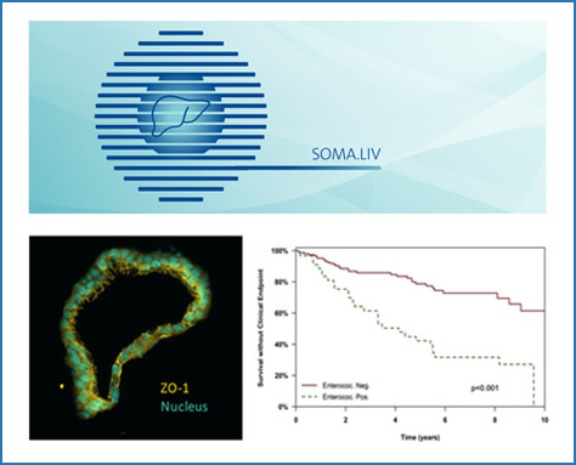
Buck L, Herzberg J, Lenaert B, Löwe B, Hartl J, Schramm C, Toussaint A. Sci Rep. 2025 Aug 26;15(1):31464. doi: 10.1038/s41598-025-16191-2. PMID: 40858902; PMCID: PMC12381113.
Abstract
Fatigue is the most common symptom in people with primary biliary cholangitis (PBC) and resistant to current treatment modalities. The aim of this study was to investigate the effect of negative and positive fatigue expectations in people with PBC on experienced fatigue and the motivational urge to stop a cognitive task. A subsample of the SOMA.LIV study of N = 46 people with PBC was randomly assigned to two experimental conditions. They received either fatigue-inducing or fatigue-reducing task instructions for a subsequent cognitive task. Participants rated their fatigue expectations prior to the task and their fatigue and motivational urge to stop after each of five task blocks. The total sample showed an increase in subjective fatigue and urge to stop across all task blocks. Participants receiving fatigue-inducing task instructions reported higher urge to stop compared to the group with fatigue-reducing task instructions. Both groups did not differ significantly in fatigue expectations and subjective fatigue. Our findings suggest that people with PBC may benefit from encouragement to engage in cognitive activities and maintain mental effort by verbal suggestions - an effect that can be of use in clinical practice to reduce potential avoidance behaviour.
ElAbd H, Pesesky M, Innocenti G, Chung BK, Mahdy AKH, Kriukova V, Kulsvehagen L, Strobbe D, Stühler C, Mayr G, May DH, Prinzensteiner M, Steiert TA, Tran F, Hadjihannas MV, Günther R, Rosati E, Mucha S, Lieb W, Ziemann M, Dempfle A, Braun F, Folseraas T, Hov JR, Melum E, Bacher P, Sterneck M, Weismüller TJ, Lenzen H, Bokemeyer B, Howie B, Robins HS, Röcken C, Schreiber S, Khanna N, Pröbstel AK, Schramm C, Vogl T, Karlsen TH, Franke A. Nat Med. 2025 Jul;31(7):2306-2316. doi: 10.1038/s41591-025-03692-w. Epub 2025 Jun 11. PMID: 40500415; PMCID: PMC12283410.
Abstract
Primary sclerosing cholangitis (PSC) is an idiopathic, progressive and incurable liver disease. Here, we aimed for systematic analyses of adaptive immune responses in PSC. By profiling the T cell repertoires of 504 individuals with PSC and 904 healthy controls, we identified 1,008 clonotypes associated with PSC. A substantial fraction of these clonotypes was restricted to known PSC human leukocyte antigen susceptibility alleles and known to target Epstein-Barr virus (EBV) epitopes. We further utilized phage-immunoprecipitation sequencing to determine antibody epitope repertoires of 120 individuals with PSC and 202 healthy controls, which showed a higher burden of anti-EBV responses in PSC than controls. EBV-specific monoclonal antibodies isolated from B cells in PSC livers corroborated convergent B and T cell responses against EBV. By analyzing electronic health records of >116 million people, we identified an association between infectious mononucleosis and PSC (odds ratio, 12; 95% confidence interval, 6.3-22.9), suggesting a link between EBV and PSC.
Henze L*, Will N*, Lee D, Haas V, Casar C, Meyer J, Stein S, Mangler F, Steinmann S, Poch T, Krause J, Fuss J, Schröder J, Kulle AE, Holterhus PM, Bonn S, Altfeld M, Huber S, Lohse AW, Schwinge D, Schramm C. JCI Insight 2025 Apr 22;10(8):e184544.
Abstract
Autoimmune hepatitis (AIH) and primary biliary cholangitis (PBC) are autoimmune liver diseases with strong female predominance. They are caused by T cell–mediated injury of hepatic parenchymal cells, but the mechanisms underlying this sex bias are unknown. Here, we investigated whether testosterone contributes to T cell activation in women with PBC. Compared with sex- and age-matched healthy controls (n = 23), cisgender (cis) women with PBC (n = 24) demonstrated decreased testosterone serum levels and proinflammatory CD4+ T cell profile in peripheral blood. Testosterone suppressed the expression of TNF and IFN-γ by human CD4+ T cells in vitro. In trans men receiving gender-affirming hormone therapy (GAHT) (n = 25), testosterone affected CD4+ T cell function by inhibiting Th1 and Th17 differentiation and by supporting the differentiation into regulatory Treg. Mechanistically, we provide evidence for a direct effect of testosterone on T cells using mice with T cell–specific deletion of the cytosolic androgen receptor. Supporting a role for testosterone in autoimmune liver disease, we observed an improved disease course and profound changes in T cell states in a trans man with AIH/primary sclerosing cholangitis (PSC) variant syndrome receiving GAHT. We here report a direct effect of testosterone on CD4+ T cells that may contribute to future personalized treatment strategies.
Holmer M, Ingre M, Färkkilä M, Ponsioen C, Mol B, Schramm C, Folseraas T, Wiencke K, Cazzagon N, Catanzaro E, Molinaro A, Nilsson E, Vessby J, Kechagias S, Nyhlin N, Werner M, Bergquist A. Hepatology 2025 Apr 16. doi: 10.1097/HEP.0000000000001351. Online ahead of print.
Abstract
Background and aims: The risk of HCC in primary sclerosing cholangitis (PSC) is unclear. Studies indicate a low risk for HCC, questioning the rationale for current HCC surveillance guidelines. This study explores the risk of HCC in a longitudinal multicenter cohort with over 3000 PSC subjects.
Approach and results: Subjects with well-characterized PSC (n=3071) were followed at 12 university hospitals within the International PSC Registry (IPSCR) collaboration for a total of 38,387 person-years. Incident HCC was registered. Subjects were followed from PSC diagnosis until death, liver transplantation, diagnosis of hepatobiliary malignancy, or February 2024. Poisson regression was used to calculate incidence rate ratios for HCC for the total population and for subgroups of different ages and cirrhosis status. Thirty-nine subjects developed HCC after a mean time of 16.4 years (SD ±10.7) from PSC diagnosis. In 26 (66.7%) of HCC cases, cirrhosis was diagnosed before HCC. The mean age at HCC diagnosis was 55.6 years (±SD13.1 years), and 28 (71.8%) were male. HCC was associated with cirrhosis (IRR: 10.8, 95% CI: 5.7-20.5) and age (IRR 1.05, 95% CI: 1.03-1.08). At the age of 50, the incidence rate was 0.81 and 0.47 for cirrhotic men and women, respectively. For non-cirrhotic subjects, the risk was low for both men and women and all age groups.
Conclusion: HCC is relatively rare in patients with primary sclerosing cholangitis who do not have cirrhosis, especially in those under the age of 50. Our findings indicate that HCC monitoring for patients with PSC can be tailored based on their age and cirrhosis status.
Werner E, van Hooff MCB, Weijsters GHX, Abbas N, Gerussi A, Willemse JA, Mitchell-Thain R, Leburgue A, Hirschfield GM, Corpechot C, Schramm C, Levy C, Nevens F, Verbeek J, Mason AL, Dalekos G, Cazzagon N, Mells GF, Kowdley KV, Carbone M, Jones DE, Hansen BE, Trivedi PJ, van der Meer AJ.Global PBC Study Group/ERN RARE-LIVER/Question Prompt List. Clin Gastroenterol Hepatol 2025 Apr 15:S1542-3565(25)00293-9. doi: 10.1016/j.cgh.2025.01.037. Online ahead of print.
Abstract
Background & aims: Clinical practice guidelines support caregivers to manage liver diseases. However, people with lifelong conditions often lack guidance to understand what aspects of care are most important and how their disease should be managed. This study aimed to create a question prompt list (QPL) for individuals with primary biliary cholangitis (PBC) including key questions (directed) to their treating physician that are most likely to improve their outcome.
Methods: International PBC professionals including patient representatives rated and ranked questions related to 9 aspects of PBC care. Questions rated by >70% as moderately/very important were considered best candidate questions (BCQs) for the QPL. Results of the survey were discussed during 2 in-person meetings, upon which the questions and/or QPL were amended.
Results: Based on 108 respondents, 11 of 43 questions were considered BCQs. After 2 in-person meetings (64 attendees), the final QPL contained 8 questions and was unanimously approved by 19 members of the study team during the consensus meeting. The included questions referred to the risk of disease progression, presence of cirrhosis, need of second-line therapy, need of repeated liver stiffness measurements, bone health, and availability of patient information and support. Two BCQs addressing options to manage pruritus and fatigue were combined on the QPL. In addition, one question regarding first-line therapy was included despite being rated as moderately/very important by 68.5%.
Conclusions: The PBC patient question prompt list serves as a user-facing document, to enhance the patient experience, and drive value-based healthcare in routine clinical practice.
Horst LJ, Zimmermann K, Lutz J, Weidemann S, Lüth S, Lohse AW, Schulze Zur Wiesch J, Schramm C, Wehmeyer MH, Müller M, Sebode M. Liver Int 2025 Mar;45(3):e70037. doi: 10.1111/liv.70037
Abstract
Background and aims: Sarcoidosis is a granulomatous multi-systemic disorder of uncertain aetiology frequently involving the liver. This study aimed to delineate the histological characteristics, treatment effectiveness and factors predictive of liver-related complications in individuals with hepatic sarcoidosis.
Methods: This retrospective cohort study included patients diagnosed with hepatic sarcoidosis by liver biopsy, which was conducted at two tertiary care centres from January 2009 to December 2023. We analysed demographic, clinical and laboratory parameters, treatment response and outcome.
Results: We enrolled 70 hepatic sarcoidosis patients with a median follow-up of 45 months (IQR 11-97 months), including 37 males with a median age of 48 years (IQR 37-59 years). Elevated GGT (94%) and ALP (81%) were the most common liver-specific biochemical alterations observed. Using mini-laparoscopy for liver biopsy made it possible to macroscopically identify granulomatous disease in 71% of patients. While at baseline, 16% of the cohort showed evidence of potential portal hypertension, at the last follow-up, 23% of patients developed complications related to portal hypertension. In addition to granulomatous changes, bile duct irregularities were found in 57% of liver biopsies, indicating cholangiopathy being part of the hepatic manifestation of sarcoidosis. Treatment with Ursodeoxycholic acid and prednisolone resulted in a significantly more pronounced decrease in ALT and ALP compared to untreated patients.
Conclusions: Patients with hepatic sarcoidosis require careful assessment of disease manifestation with a particular focus on portal hypertension. Treatment with UDCA and prednisolone leads to a reduction of biochemical parameters in a significant proportion of these patients.
Wang SH, Serr I, Digigow R, Metzler B, Surnov A, Gottwick C, Alsamman M, Krzikalla D, Heine M, Zahlten M, Widera A, Mungalpara D, Şeleci M, Fanzutti M, Marques Mesquita LM, Vocaturo AL, Herkel J, Carambia A, Schröter C, Sarko D, Pohlner J, Daniel C, de Min C, Fleischer S. Front Immunol. 2025 Mar 17;16:1542380. doi: 10.3389/fimmu.2025.1542380. PMID: 40165970; PMCID: PMC11955608.
Abstract
Introduction: Treating autoimmune diseases without nonspecific immunosuppression remains challenging. To prevent or treat these conditions through targeted immunotherapy, we developed a clinical-stage nanoparticle platform that leverages the tolerogenic capacity of liver sinusoidal endothelial cells (LSECs) to restore antigen-specific immune tolerance.
Methods: In vivo efficacy was evaluated in various CD4+ T cell-mediated disease models, including preventive and therapeutic models of myelin oligodendrocyte glycoprotein-induced experimental autoimmune encephalomyelitis (EAE), ovalbumin-sensitized delayed-type hypersensitivity (DTH), and the spontaneous type 1 diabetes model. Nanoparticle-induced antigen-specific immune responses were also analyzed through adoptive transfers of 2D2 transgenic T cells into wild-type mice, followed by nanoparticle administration.
Results: The peptide-conjugated nanoparticles displayed a uniform size distribution (25–30 nm). Their coupling efficiency for peptides with unfavorable physicochemical properties was significantly enhanced by a proprietary linker technology. Preferential LSEC targeting of nanoparticles coupled with fluorescently labeled peptides was confirmed via intravital microscopy and flow cytometry. Intravenous nanoparticle administration significantly reduced disease severity and demyelination in EAE, independent of prednisone at maintenance doses, and suppressed target tissue inflammation in the DTH model. Furthermore, prophylactic administration of a mixture of nanoparticles coupled with five autoantigenic peptides significantly lowered the hyperglycemia incidence of the non-obese diabetic mice. Mechanistically, the tolerizing effects were associated with the induction of antigen-specific regulatory T cells and T cell anergy, which counteract proinflammatory T cells in the target tissue.
Conclusion: Our findings demonstrate that peptide-loaded nanoparticles preferentially deliver disease-relevant peptides to LSECs, thereby inducing antigen-specific immune tolerance. This versatile clinical-stage nanoparticle platform holds promise for clinical application across multiple autoimmune diseases.
Snijders RJALM, Janik MK, Mund M, Uhlenbusch N, Raszeja-Wyszomirska J, Gerussi A, Bolis F, Cristoferi L, Invernizzi P, Kovats P, Papp M, Grønbæk L, Grønbæk H, Tjwa ETTL, Aamann L, Ytting H, Ronca V, Olsen K, Oo YH, van der Meer AJ, Madaleno J, Canhão B, Engel B, Campos-Murguia A, Taubert R, Koc ÖM, Kramer M, Willemse JA, Löwe B, Lohse AW, Drenth JPH, Schramm C, Milkiewicz P, Gevers TJ. Hepatology 2025 Feb 19. doi: 10.1097/HEP.0000000000001271. Online ahead of print
Abstract
Background and aims: Impaired health-related quality of life (HRQoL) contributes to the overall disease burden in autoimmune hepatitis (AIH). This study aimed to evaluate HRQoL in people with AIH and to identify potentially modifiable factors associated with impaired HRQoL using validated patient-reported outcome measures.
Approach and results: Adult patients with AIH diagnosed at 12 European centers were enrolled in this prospective, cross-sectional study from July 2020 to June 2023. HRQoL was assessed using the Physical Component Score (PCS) and Mental Component Score (MCS) of the 12-item Short Form Health Survey (SF-12), and the European Quality-of-life 5-Dimension 5-Level (EQ-5D-5L) utility index score. Mixed-model regression analyses identified factors associated with HRQoL and somatic symptom severity. Controls were recruited from the general population in 5 European countries. A total of 882 patients with AIH (mean age: 51.0 y [SD: 17.0]; 76.4% female) and 178 controls were included. Physical but not mental HRQoL was impaired in the AIH group compared with the control group (PCS: 46.3 vs. 51.9, p <0.001; EQ-5D utility index score: 0.87 vs. 0.95, p <0.001). HRQoL was associated with severe somatic symptoms (PCS: β=-4.26, p <0.001), fatigue (PCS: β=-0.25, p <0.001; MCS: β=-0.25, p <0.001), and depression/anxiety (PCS: β=-3.37, p <0.001; MCS: β=-6.79, p <0.001). A complete biochemical response was associated with lower somatic symptom severity (OR: 0.69, p <0.05).
Conclusions: People with AIH had impaired HRQoL compared with controls, particularly in terms of physical well-being. HRQoL scores are associated with symptom burden, encompassing both somatic and psychosocial dimensions.
Gronke K, Nguyen M, Fuhrmann H, Santamaria de Souza N, Schumacher J, Pereira MS, Löschberger U, Brinkhege A, Becker NJ, Yang Y, Sonnert N, Leopold S, Martin AL, von Münchow-Klein L, Pessoa Rodrigues C, Cansever D, Hallet R, Richter K, Schubert DA, Daniel GM, Dylus D, Forkel M, Schwinge D, Schramm C, Redanz S, Lassen KG, Manfredo Vieira S, Piali L, Palm NW, Bieniossek C, Kriegel MA. Sci Transl Med 2025 Feb 5;17(784):eadj6294. doi: 10.1126/scitranslmed.adj6294. Epub 2025 Feb 5.PMID: 39908347
Abstract
Chronic autoimmune diseases often lead to long-term sequelae and require lifelong immunosuppression because of an incomplete understanding of the triggers and drivers in genetically predisposed patients. Gut bacteria that escape the gut barrier, known as translocating gut pathobionts, have been implicated as instigators and perpetuators of extraintestinal autoimmune diseases in mice. The gut microbial contributions to autoimmunity in humans remain largely unclear, including whether specific pathological human adaptive immune responses are triggered by such pathobionts. Here, we show that the translocating pathobiont Enterococcus gallinarum can induce both human and mouse interferon-γ+ T helper 17 (TH17) differentiation and immunoglobulin G3 (IgG3) subclass switch of anti-E. gallinarum RNA antibodies, which correlated with anti-human RNA autoantibody responses in patients with systemic lupus erythematosus (SLE) and autoimmune hepatitis, two extraintestinal autoimmune diseases. E. gallinarum RNA, but not human RNA, triggered Toll-like receptor 8 (TLR8), and TLR8-mediated human monocyte activation promoted human TH17 induction by E. gallinarum. Translocation of the pathobiont triggered increased anti-RNA autoantibody titers that correlated with renal autoimmune pathophysiology in murine gnotobiotic lupus models and with disease activity in patients with SLE. These studies elucidate cellular mechanisms of how a translocating gut pathobiont induces systemic human T cell- and B cell-dependent autoimmune responses and provide a framework for developing host- and microbiota-derived biomarkers and targeted therapies in autoimmune diseases.
Horst LJ, Kempski J, Walmsley M, Huber S, Schramm C. Hepatology 2025 Jan 22.
Abstract
Primary sclerosing cholangitis is one of the most challenging conditions in hepatology, and due to our limited understanding of its pathogenesis, no causal therapies are currently available. While it was long assumed that a minority of people with inflammatory bowel disease (IBD) also develop primary sclerosing cholangitis (PSC), which is sometimes labeled an extraintestinal manifestation of IBD, the clinical phenotype, genetic, and intestinal microbiota associations strongly argue for PSC-IBD being a distinct form of IBD, existing alongside ulcerative colitis and Crohn’s disease. In fact, the liver itself could contribute to intestinal pathology, clinically overt in 60%–80% of patients. Recent studies suggested that on a molecular level, almost all people with PSC have underlying colitis. The extent to which the liver and gut influence each other clinically and in terms of disease progression has not yet been conclusively revealed. However, while it seemed intuitive that the 2 diseases have a negative influence on each other, evidence suggests that sclerosing cholangitis can also be protective for the gut and that colitis can, in certain settings, ameliorate liver pathology. This underscores the complex pathophysiological relationships, where factors such as genetic predisposition, changes in the intestinal microbiota, altered bile acid metabolism, and immune cell migration are among the suspected contributors. PSC is an emerging disease with a significant impact on the health-related quality of life of affected people. With this review, we aim to summarize the current knowledge on the gut-liver axis in PSC-IBD, provide new perspectives on risk stratification and treatment, and identify gaps in our current knowledge. Our understanding of this complex relationship will therefore help to design clinical trials and shape the future therapy of PSC-IBD.
Publications 2024
Laschtowitz A, Lindberg EL, Liebhoff AM, Liebig LA, Casar C, Steinmann S, Guillot A, Xu J, Schwinge D, Trauner M, Lohse AW, Bonn S, Hübner N, Schramm C. JHEP Rep 2024 Nov 12;7(3):101267.
Abstract
Background & aims: Primary sclerosing cholangitis (PSC) is a chronic heterogenous cholangiopathy with unknown etiology where chronic inflammation of the bile ducts leads to multifocal biliary strictures and biliary fibrosis with consecutive cirrhosis development. We here aimed to identify a PSC-specific gene signature associated with biliary fibrosis development.
Methods: We performed RNA-sequencing of 47 liver biopsies from people with PSC (n = 16), primary biliary cholangitis (PBC, n = 15), and metabolic dysfunction-associated steatotic liver disease (MASLD, n = 16) with different fibrosis stages to identify a PSC-specific gene signature associated with biliary fibrosis progression. For validation, we compared an external transcriptome data set of liver biopsies from people with PSC (n = 73) with different fibrosis stages (baseline samples from NCT01672853).
Results: Differential gene expression analysis of the liver transcriptome from patients with PSC with advanced vs. early fibrosis revealed 431 genes associated with fibrosis development. Of those, 367 were identified as PSC-associated when compared with PBC or MASLD. Validation against an external data set of 73 liver biopsies from patients with PSC with different fibrosis stages led to a condensed set of 150 (out of 367) differentially expressed genes. Cell type specificity assignment of those genes by using published single-cell RNA-Seq data revealed genetic disease drivers expressed by cholangiocytes (e.g. CXCL1, SPP1), fibroblasts, innate, and adaptive immune cells while deconvolution along fibrosis progression of the PSC, PBC, and MASLD samples highlighted an early involvement of macrophage- and neutrophil-associated genes in PSC fibrosis.
Conclusions: We reveal a PSC-attributed gene signature associated with biliary fibrosis development that may enable the identification of potential new biomarkers and therapeutic targets in PSC-related fibrogenesis.
Impact and implications: Primary sclerosing cholangitis (PSC) is an inflammatory liver disease that is characterized by multifocal inflammation of bile ducts and subsequent biliary fibrosis. Herein, we identify a PSC-specific gene set of biliary fibrosis progression attributing to a uniquely complex milieu of different cell types, including innate and adaptive immune cells while neutrophils and macrophages showed an earlier involvement in fibrosis initiation in PSC in contrast to PBC and metabolic dysfunction-associated steatotic liver disease. Thus, our unbiased approach lays an important groundwork for further mechanistic studies for research into PSC-specific fibrosis.
Liwinski T*, Auer MK*, Schröder J, Pieknik I, Casar C, Schwinge D, Henze L, Stalla GK, Lang UE, von Klitzing A, Briken P, Hildebrandt T, Desbuleux JC, Biedermann SV, Holterhus P-M, Bang C, Schramm C*, Fuss J*. BMC Medicine 2024;22:346.
Abstract
Background: Limited data exists regarding gender-specific microbial alterations during gender-affirming hormonal therapy (GAHT) in transgender individuals. This study aimed to investigate the nuanced impact of sex steroids on gut microbiota taxonomy and function, addressing this gap. We prospectively analyzed gut metagenome changes associated with 12 weeks of GAHT in trans women and trans men, examining both taxonomic and functional shifts.
Methods: Thirty-six transgender individuals (17 trans women, 19 trans men) provided pre- and post-GAHT stool samples. Shotgun metagenomic sequencing was used to assess the changes in gut microbiota structure and potential function following GAHT.
Results: While alpha and beta diversity remained unchanged during transition, specific species, including Parabacteroides goldsteinii and Escherichia coli, exhibited significant abundance shifts aligned with affirmed gender. Overall functional metagenome analysis showed a statistically significant effect of gender and transition (R2 = 4.1%, P = 0.0115), emphasizing transitions aligned with affirmed gender, particularly in fatty acid-related metabolism.
Conclusions: This study provides compelling evidence of distinct taxonomic and functional profiles in the gut microbiota between trans men and women. GAHT induces androgenization in trans men and feminization in trans women, potentially impacting physiological and health-related outcomes.
Hartl J, Buck L, Löwe B, Toussaint A, Schramm C. J Hepatol 2025;82:e44-e45.
Abstract
Although fatigue has been identified as a major determinant of health-related quality of life (HRQL) in chronic liver disease (CLD), it has received only little attention from scientists or physicians in the past. Therefore, we are pleased that fatigue has been given the spotlight in a recent review article by Younossi et al. in the Journal of Hepatology. As highlighted by the authors, while improvement or cure in certain CLDs may alleviate fatigue, currently licensed treatment options do not improve fatigue in primary biliary cholangitis (PBC), which represents the CLD in which fatigue has the greatest impact on HRQL, affecting 30 – 60% of patients. Therefore, investigating fatigue in PBC addresses a major unmet clinical need for patients. This should not narrow our clinical focus to PBC and distract from the fact that fatigue can also be relevant and severe in other autoimmune liver diseases such as primary sclerosing cholangitis (PSC). However, as pointed out in the current review, data on fatigue from large cohorts of patients with PSC are lacking so far.
Therefore, we prospectively explored the frequency and severity of fatigue, as well as potential contributing biomedical factors in a large, well-characterized cohort of patients with PSC (n = 228) compared to patients with PBC (n = 238), according to the previously published study protocol (SOMA.LIV; trial registration number ISRCTN14379650). Fatigue was assessed by PBC-40, a patient-derived, disease-specific quality of life measure with robust psychometric properties, including a fatigue domain that is also commonly used as patient-reported outcome measure in PSC.
In PBC, 29% of the patients had a mean PBC-40 fatigue domain score greater than 33, which is considered clinically significant fatigue. Fatigue severity correlated with younger age at diagnosis (r = -0.20, p = 0.004), but not with any biochemical markers, such as alkaline phosphatase, or stage of liver disease as assessed by liver stiffness. Of note, when we explored the sex impact, questionnaires revealed a significantly higher median fatigue score among women than in men (26 vs. 17 [median], p = 0.003;
Poch T*, Bahn J*, Casar C, Krause J, Evangelakos I, Gilladi H, Kunzmann LK, Laschtowitz A, Iuso N, Schäfer AM, Liebig LA, Steinmann S, Sebode M, Folseraas T, Engesæter LK, Karlsen TH, Franke A, Hubner N, Schlein C, Galun E, Huber S, Lohse AW, Gagliani N, Schwinge D*, Schramm C*. Cell Rep Med 2024 Jun 13:101620. doi: 10.1016/j.xcrm.2024.101620. Online ahead of print. PMID: 38901430
Abstract
Primary sclerosing cholangitis (PSC) is an immune-mediated liver disease of unknown pathogenesis, with a high risk to develop cirrhosis and malignancies. Functional dysregulation of T cells and association with genetic polymorphisms in T cell-related genes were previously reported for PSC. Here, we genotyped a representative PSC cohort for several disease-associated risk loci and identified rs56258221 (BACH2/MIR4464) to correlate with not only the peripheral blood T cell immunophenotype but also the functional capacities of naive CD4+ T (CD4+ TN) cells in people with PSC. Mechanistically, rs56258221 leads to an increased expression of miR4464, in turn causing attenuated translation of BACH2, a major gatekeeper of T cell quiescence. Thereby, the fate of CD4+TN is skewed toward polarization into pro-inflammatory subsets. Clinically, people with PSC carrying rs56258221 show signs of accelerated disease progression. The data presented here highlight the importance of assigning functional outcomes to disease-associated genetic polymorphisms as potential drivers of diseases.
Weltzsch JP, Bartel CF, Waldmann M, Renné T, Schulze S, Terziroli Beretta-Piccoli B, Papp M, Ye O, Ronca V, Sebode M, Lohse AW, Schramm C, Hartl J. Hepatology 2024;80:1026-1040.
Abstract
Background and aims: In autoimmune hepatitis, achieving complete biochemical remission (CBR) with current weight-based thiopurine dosing is challenging. We investigated whether patients could be stratified regarding CBR according to a target range of thiopurine metabolites. Moreover, we explored the effects of azathioprine dosage increases and co-therapy of allopurinol with low-dose thiopurines on metabolite profiles and treatment response.
Approach and results: The relation between metabolites and treatment response was assessed in 337 individuals from 4 European centers. In a global, cross-sectional analysis, active metabolites 6-thioguanine nucleotides (6TGN) were similar in those with and without CBR. However, analyzing patients with sequential measurements over 4 years (N = 146) revealed higher average 6TGN levels in those with stable CBR (260 pmol/0.2 mL) compared to those failing to maintain CBR (181 pmol/0.2 mL; p = 0.0014) or never achieving CBR (153 pmol/0.2 mL; p < 0.0001), with an optimal 6TGN cutoff of ≥223 pmol/0.2 mL (sensitivity: 76% and specificity: 78%). Only 42% exhibited 6TGN ≥223 pmol/0.2 mL following weight-based dosing, as doses weakly correlated with 6TGN but with 6-methylmercaptopurine (6MMP), a metabolite associated with toxicity. Azathioprine dose increases led to preferential 6MMP formation (+127% vs. 6TGN +34%; p < 0.0001). Conversely, adding allopurinol to thiopurines in difficult-to-treat patients (N = 36) raised 6TGN (168→321 pmol/0.2 mL; p < 0.0001) and lowered 6MMP (2125→184 pmol/0.2 mL; p < 0.0001), resulting in improved transaminases in all patients and long-term CBR in 75%.
Conclusions: Maintaining CBR in autoimmune hepatitis was associated with 6TGN ≥223 pmol/0.2 mL. For patients who fail to achieve CBR and therapeutic 6TGN levels despite thiopurine dose increase due to preferential 6MMP formation, comedication of allopurinol alongside low-dose thiopurines represents an efficient alternative.
Zecher BF, Ellinghaus D, Schloer S, Niehrs A, Padoan B, Baumdick ME, Yuki Y, Martin MP, Glow D, Schröder-Schwarz J, Niersch J, Brias S, Müller LM, Habermann R, Kretschmer P, Früh T, Dänekas J, Wehmeyer MH, Poch T, Sebode M; International PSC Study Group (IPSCSG); Ellinghaus E, Degenhardt F, Körner C, Hoelzemer A, Fehse B, Oldhafer KJ, Schumacher U, Sauter G, Carrington M, Franke A, Bunders MJ, Schramm C*, Altfeld M*. Gut 2024 Jan 5;73(2):325-337.
Abstract
Objective: Primary sclerosing cholangitis (PSC) is characterised by bile duct strictures and progressive liver disease, eventually requiring liver transplantation. Although the pathogenesis of PSC remains incompletely understood, strong associations with HLA-class II haplotypes have been described. As specific HLA-DP molecules can bind the activating NK-cell receptor NKp44, we investigated the role of HLA-DP/NKp44-interactions in PSC.
Design: Liver tissue, intrahepatic and peripheral blood lymphocytes of individuals with PSC and control individuals were characterised using flow cytometry, immunohistochemical and immunofluorescence analyses. HLA-DPA1 and HLA-DPB1 imputation and association analyses were performed in 3408 individuals with PSC and 34 213 controls. NK cell activation on NKp44/HLA-DP interactions was assessed in vitro using plate-bound HLA-DP molecules and HLA-DPB wildtype versus knock-out human cholangiocyte organoids.
Results: NKp44+NK cells were enriched in livers, and intrahepatic bile ducts of individuals with PSC showed higher expression of HLA-DP. HLA-DP haplotype analysis revealed a highly elevated PSC risk for HLA-DPA1*02:01~B1*01:01 (OR 1.99, p=6.7×10−50). Primary NKp44+NK cells exhibited significantly higher degranulation in response to plate-bound HLA-DPA1*02:01-DPB1*01:01 compared with control HLA-DP molecules, which were inhibited by anti-NKp44-blocking. Human cholangiocyte organoids expressing HLA-DPA1*02:01-DPB1*01:01 after IFN-γ-exposure demonstrated significantly increased binding to NKp44-Fc constructs compared with unstimulated controls. Importantly, HLA-DPA1*02:01-DPB1*01:01-expressing organoids increased degranulation of NKp44+NK cells compared with HLA-DPB1-KO organoids.
Conclusion: Our studies identify a novel PSC risk haplotype HLA-DP A1*02:01~DPB1*01:01 and provide clinical and functional data implicating NKp44+NK cells that recognise HLA-DPA1*02:01-DPB1*01:01 expressed on cholangiocytes in PSC pathogenesis.
Publications 2023
Cell Mol Gastroenterol Hepatol. Krzikalla D, Laschtowitz A, Leypoldt L, Gottwick C, Averhoff P, Weidemann S, Lohse AW, Huber S, Schramm C, Schwinge D, Herkel J, Carambia A. IFNγ and CTLA-4 Drive 2024;17(1):79-91.
Abstract
Background & aims: The liver has a distinct capacity to induce immune tolerance to hepatic antigens. Although liver tolerance can be advantageous for preventing autoimmune and inflammatory diseases, it also can be detrimental by preventing immune surveillance of infected or malignant cells. Here, we investigated the immune mechanisms that establish hepatic tolerance.
Methods: Tolerance was investigated in C-reactive protein (CRP)-myelin basic protein (MBP) mice expressing the neuroantigen MBP in hepatocytes, providing profound resistance to MBP-induced neuroinflammation. Tolerance induction was studied after transfer of MBP-specific CD4 T cells into CRP-MBP mice, and tolerance mechanisms were tested using depleting or blocking antibodies.
Results: Although tolerant CRP-MBP mice display increased numbers of forkhead box P3+ regulatory T cells, we here found them not essential for the maintenance of hepatic tolerance. Instead, upon MBP recognition in the liver, MBP-specific T cells became activated to produce interferon (IFN)γ, which, in turn, induced local up-regulation of recruitment molecules, including Chemokine (C-X-C motif) ligand9 and its receptor C-X-C motif chemokine receptor3, facilitating endothelial translocation and redirection of MBP-specific T cells into the hepatic parenchyma. There, the translocated MBP-specific CD4 T cells partly converted into interleukin 10-producing type 1 regulatory T cells, and significantly up-regulated the expression of immune checkpoint molecules, notably cytotoxic T-lymphocyte-associated protein 4 (CTLA-4). Intriguingly, although liver tolerance was not affected by impairment of interleukin 10 signaling, concomitant blockade of IFNγ and CTLA-4 abrogated hepatic tolerance induction to MBP, resulting in neuroinflammatory autoimmune disease in these mice.
Conclusions: IFNγ-mediated redirection of autoreactive CD4 T cells into the liver and up-regulation of checkpoint molecules, including CTLA-4, were essential for tolerance induction in the liver, hence representing a potential treatment target for boosting or preventing liver tolerance.
Abstract
During metastasis, cancer cells invade, intravasate, enter the circulation, extravasate, and colonize target organs. Here, we examined the role of interleukin (IL)-22 in metastasis. Immune cell-derived IL-22 acts on epithelial tissues, promoting regeneration and healing upon tissue damage, but it is also associated with malignancy. Il22-deficient mice and mice treated with an IL-22 antibody were protected from colon-cancer-derived liver and lung metastasis formation, while overexpression of IL-22 promoted metastasis. Mechanistically, IL-22 acted on endothelial cells, promoting endothelial permeability and cancer cell transmigration via induction of endothelial aminopeptidase N. Multi-parameter flow cytometry and single-cell sequencing of immune cells isolated during cancer cell extravasation into the liver revealed iNKT17 cells as source of IL-22. iNKT-cell-deficient mice exhibited reduced metastases, which was reversed by injection of wild type, but not Il22-deficient, invariant natural killer T (iNKT) cells. IL-22-producing iNKT cells promoting metastasis were tissue resident, as demonstrated by parabiosis. Thus, IL-22 may present a therapeutic target for prevention of metastasis.
Previous publications
Müller AL, Casar C, Preti M, Krzikalla D, Gottwick C, Averhoff P, Rosenstiel P, Gelderblom M, Altfeld M, Lohse AW, Steinmann S, Sebode M, Krause J, Schwinge D, Schramm C, Carambia A, Herkel J. J Hepatol 2022 Jul 4:S0168-8278(22)02923-3. doi: 10.1016/j.jhep.2022.06.025.
Lay summary
Primary sclerosing cholangitis (PSC) is an inflammatory liver disease of the bile ducts for which there is no effective treatment. Herein, we show that the inflammatory immune response to bile duct injury is organised by a specific subtype of immune cell called conventional type 2 dendritic cells. Our findings suggest that this cell subtype and the inflammatory molecules it produces are potential therapeutic targets for PSC.
Poch T#, Krause J#, Casar C, Liwinski T, Glau L, Kaufmann M, Ahrenstorf AE, Hess LU, Ziegler AE, Martrus G, Lunemann S, Sebode M, Li J, Schwinge D, Krebs CF, Franke A, Friese MA, Oldhafer KJ, Fischer L, Altfeld M, Lohse AW, Huber S, Tolosa E#, Gagliani N #, Schramm C #. J Hepatol 2021;75:414-423.
Lay summary
The composition of intrahepatic immune cells in primary sclerosing cholangitis (PSC) and their contribution to disease pathogenesis is widely unknown. We analysed intrahepatic T cells and identified a previously uncharacterized population of liver-resident CD4+ T cells which are expanded in the livers of patients with PSC compared to healthy liver tissue and other liver diseases. These cells are likely to contribute to the pathogenesis of PSC and could be targeted in novel therapeutic approaches.
Stein S, Henze L, Poch T, Carambia A, Krech T, Preti M, Schuran FA, Reich M, Keitel V, Fiorotto R, Strazzabosco M, Fischer L, Li J, Müller LM, Wagner J, Gagliani N, Herkel J, Schwinge D, Schramm C. J Hepatol 2021;74:919-930. doi: 10.1016/j.jhep.2020.10.035. Epub 2020 Nov 13.
Lay summary
IL-17 is assumed to be a driver of inflammation in several autoimmune diseases, such as psoriasis. IL-17 is also present in inflammatory diseases of the bile duct, but its role in these conditions is not clear, as the effects of IL-17 depend on the context of its expression. Herein, we investigated the role of IL-17 in an experimental autoimmune cholangitis mouse model, and we identified an important protective effect of IL-17 on cholangiocytes, enabling them to downregulate bile duct inflammation via checkpoint inhibitor PD-L1.
Liwinski T*, Zenouzi R*, John C, Ehlken H, Rühlemann MC, Bang C, Groth S, Lieb W, Kantowski M, Andersen N, Schachschal G, Karlsen TH, Hov JR, Rösch T, Lohse AW, Heeren J, Franke A, Schramm C. Gut 2020;69:665-672. doi: 10.1136/gutjnl-2019-318416. Epub 2019 Jun 26.
Abstract
Background: Patients with primary sclerosing cholangitis (PSC) display an altered colonic microbiome compared with healthy controls. However, little is known on the bile duct microbiome and its interplay with bile acid metabolism in PSC.
Methods: Patients with PSC (n=43) and controls without sclerosing cholangitis (n=22) requiring endoscopic retrograde cholangiography were included prospectively. Leading indications in controls were sporadic choledocholithiasis and papillary adenoma. A total of 260 biospecimens were collected from the oral cavity, duodenal fluid and mucosa and ductal bile. Microbiomes of the upper alimentary tract and ductal bile were profiled by sequencing the 16S-rRNA-encoding gene (V1–V2). Bile fluid bile acid composition was measured by high-performance liquid chromatography mass spectrometry and validated in an external cohort (n=20).
Results: The bile fluid harboured a diverse microbiome that was distinct from the oral cavity, the duodenal fluid and duodenal mucosa communities. The upper alimentary tract microbiome differed between PSC patients and controls. However, the strongest differences between PSC patients and controls were observed in the ductal bile fluid, including reduced biodiversity (Shannon entropy, p=0.0127) and increase of pathogen Enterococcus faecalis (FDR=4.18×10⁻⁵) in PSC. Enterococcus abundance in ductal bile was strongly correlated with concentration of the noxious secondary bile acid taurolithocholic acid (r=0.60, p=0.0021).
Conclusion: PSC is characterised by an altered microbiome of the upper alimentary tract and bile ducts. Biliary dysbiosis is linked with increased concentrations of the proinflammatory and potentially cancerogenic agent taurolithocholic acid.
Team


Prof. Dr. med. Ansgar Lohse
Principal Investigator
E-mail address:


Prof. Dr. med. Christoph Schramm
Principal Investigator
E-mail address:

Prof. Dr. rer. nat. Johannes Herkel
Principal Investigator
E-mail address: