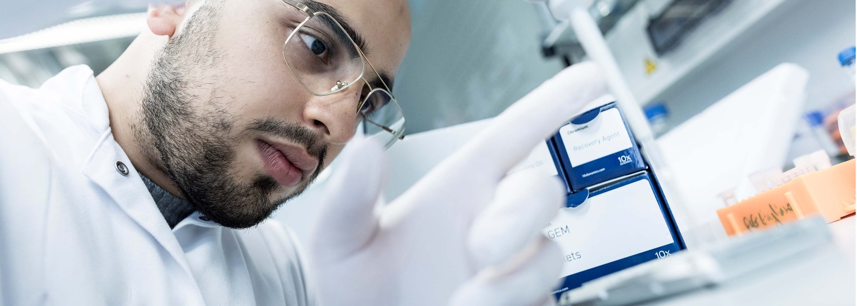

T cell biology, immunity, and immune-mediated inflammatory diseases
We like studying the basic molecular and cellular mechanisms of T cell biology, adaptation to environmental changes, and ultimately, how T cells
either mediate physiological responses to pathogens or pathological tissue damaging processes.

“Unraveling the complexities of CD4+ T cell biology in immune-related disorders.”
Prof. Dr. Nicola Gagliani
Project details and goals
Our laboratory focuses on unraveling the complexities of CD4 T cell biology, in particular their metabolism, adaptability, and roles in disease.
We investigate the different CD4 T cell subsets by exploring their developmental trajectories and the molecular mechanisms underlying their adaptability.
A key aspect of our research is understanding how CD4 T cells adapt to environmental changes, including both classical immune challenges like infections and non-classical stimuli such as dietary shifts.
We are particularly interested in the organ-specific responses of resident CD4 T cells and their implications for immune adaptation. Finally, we study the involvement of CD4 T cells in immune-mediated inflammatory diseases and cancer, profiling their interactions with other immune cells, for example macrophages, and with the different tissues at a single-cell level.
Through these efforts, we aim to contribute to a deeper understanding of CD4 T cell function and their potential in treating immune-related disorders.
Current projects
Basic Aspects of CD4+ T Cell Biology
CD4+ T cells are a plastic and heterogeneous population made up of subsets that differ in cytokine production, cell surface markers, function and tissue specificity. Although there have been efforts to classify these subsets based on surface markers and signature cytokines, unsupervised analysis of CD4+ T cell populations shows an unexpected heterogeneity which is not reflected by the classical textbook nomenclature. Supporting this, the use of cytokine fate-mapping mouse models revealed that some CD4+ T cell subsets can adapt their functionality during the development of the immune response. Redefining the classification of CD4+ T cells by understanding their plastic developmental trajectories and identifying the epigenetic and molecular mechanisms controlling these trajectories are some of the main topics of our group.

Immune adaptation of CD4+ T cells to environmental changes
CD4+ T cells possess the ability to switch from a pro- to an anti-inflammatory function, i.e. immune adaptation. The consequences of dysfunctional immune adaptation are clear: the immune cells either fail to mount a protective response to lethal infections, or they overreact to non-harmful antigens and, in doing so, induce a variety of immune-mediated inflammatory diseases (Xu H. et al Nat Comm 2020).
CD4+ T cells adapt in response to both “classical” immune stimuli, such as infections, and “non-classical” immune stimuli, such as changes in dietary habits. In addition, the immune adaptation of CD4+ T cells varies across different organs. There is indeed a conspicuous number of resident CD4+ T cells that have established an intimate and diverse relationship with each organ, which is not yet fully understood.
Our lab is particularly interested in decoding the organ-specific response of resident CD4+ T cells to different classical and non-classical stimuli, with a particular focus on the diet.

The role of CD4+ T cells in immune-mediated inflammatory diseases and cancer
CD4+ T cells are key players in orchestrating a context-appropriate immune response, but their protection comes at a price. They can, in fact, also promote immune-mediated inflammatory diseases (IMIDs), including the development of inflammation-induced carcinogenesis. By profiling resident T cells and their interactions with other immune and non-immune cells in a broad spectrum of diseased human organs at single-cell resolution, we are delineating a new anthology of T cell function in IMIDs and cancer. Using in vitro 3D culture, we validate the newly discovered functions of these cells and provide the scientific and clinical community with new targets for potential future immune therapies.
Publications 2024
Liebold I, Al Jawazneh A, Casar C, Lanzloth C, Leyk S, Hamley M, Wong MN, Kylies D, Gräfe SK, Edenhofer I, Aranda-Pardos I, Kriwet M, Haas H, Krause J, Hadjilaou A, Schromm AB, Richardt U, Eggert P, Tappe D, Weidemann SA, Ghosh S, Krebs CF, A-Gonzalez N, Worthmann A, Lohse AW, Huber S, Rothlin CV, Puelles VG, Jacobs T, Gagliani N, Bosurgi L. Science. 2024 Apr 5;384(6691):eabo7027.
Abstract
Macrophages are functionally heterogeneous cells essential for apoptotic cell clearance. Apoptotic cells are defined by homogeneous characteristics, ignoring their original cell lineage identity. We found that in an interleukin-4 (IL-4)-enriched environment, the sensing of apoptotic neutrophils by macrophages triggered their tissue remodeling signature. Engulfment of apoptotic hepatocytes promoted a tolerogenic phenotype, whereas phagocytosis of T cells had little effect on IL-4-induced gene expression. In a mouse model of parasite-induced pathology, the transfer of macrophages conditioned with IL-4 and apoptotic neutrophils promoted parasitic egg clearance. Knockout of phagocytic receptors required for the uptake of apoptotic neutrophils and partially T cells, but not hepatocytes, exacerbated helminth infection. These findings suggest that the identity of apoptotic cells may contribute to the development of distinct IL-4-driven immune programs in macrophages.
Publications 2023
Abstract
Omnivorous animals, including mice and humans, tend to prefer energy-dense nutrients rich in fat over plant-based diets, especially for short periods of time, but the health consequences of this short-term consumption of energy-dense nutrients are unclear. Here, we show that short-term reiterative switching to 'feast diets', mimicking our social eating behavior, breaches the potential buffering effect of the intestinal microbiota and reorganizes the immunological architecture of mucosa-associated lymphoid tissues. The first dietary switch was sufficient to induce transient mucosal immune depression and suppress systemic immunity, leading to higher susceptibility to Salmonella enterica serovar Typhimurium and Listeria monocytogenes infections. The ability to respond to antigenic challenges with a model antigen was also impaired. These observations could be explained by a reduction of CD4+ T cell metabolic fitness and cytokine production due to impaired mTOR activity in response to reduced microbial provision of fiber metabolites. Reintroducing dietary fiber rewired T cell metabolism and restored mucosal and systemic CD4+ T cell functions and immunity. Finally, dietary intervention with human volunteers confirmed the effect of short-term dietary switches on human CD4+ T cell functionality. Therefore, short-term nutritional changes cause a transient depression of mucosal and systemic immunity, creating a window of opportunity for pathogenic infection.
Abstract
Pancreatic ductal adenocarcinoma (PDAC) is expected to be the second most deadly cancer by 2040, owing to the high incidence of metastatic disease and limited responses to treatment. Less than half of all patients respond to the primary treatment for PDAC, chemotherapy, and genetic alterations alone cannot explain this. Diet is an environmental factor that can influence the response to therapies, but its role in PDAC is unclear. Here, using shotgun metagenomic sequencing and metabolomic screening, we show that the microbiota-derived tryptophan metabolite indole-3-acetic acid (3-IAA) is enriched in patients who respond to treatment. Faecal microbiota transplantation, short-term dietary manipulation of tryptophan and oral 3-IAA administration increase the efficacy of chemotherapy in humanized gnotobiotic mouse models of PDAC. Using a combination of loss- and gain-of-function experiments, we show that the efficacy of 3-IAA and chemotherapy is licensed by neutrophil-derived myeloperoxidase. Myeloperoxidase oxidizes 3-IAA, which in combination with chemotherapy induces a downregulation of the reactive oxygen species (ROS)-degrading enzymes glutathione peroxidase 3 and glutathione peroxidase 7. All of this results in the accumulation of ROS and the downregulation of autophagy in cancer cells, which compromises their metabolic fitness and, ultimately, their proliferation. In humans, we observed a significant correlation between the levels of 3-IAA and the efficacy of therapy in two independent PDAC cohorts. In summary, we identify a microbiota-derived metabolite that has clinical implications in the treatment of PDAC, and provide a motivation for considering nutritional interventions during the treatment of patients with cancer.
Previous publications
Lilan Zhao*, Anastasios D Giannou*, Yang Xu*, Ahmad Mustafa Shiri, Imke Liebold, Babett Steglich, Tanja Bedke, Tao Zhang, Jöran Lücke, Pasquale Scognamiglio, Jan Kempski, Anna Woestemeier, Jing Chen, Theodora Agalioti, Dimitra E Zazara, Diana Lindner, Melanie Janning, Jan K Hennigs, Rajesh M Jagirdar, Ourania S Kotsiou, Sotirios G Zarogiannis, Yasushi Kobayashi, Jacob R Izbicki, Sourav Ghosh, Carla V Rothlin, Lidia Bosurgi#, Samuel Huber#, Nicola Gagliani#
*These authors contributed equally to this work
# These authors cosuprevised equally to this work
Science Adv. 2021 Aug 13;7(33):eabd6734
Abstract
Malignant pleural effusion (MPE) results from the capacity of several human cancers to metastasize to the pleural cavity. No effective treatments are currently available, reflecting our insufficient understanding of the basic mechanisms leading to MPE progression. Here, we found that efferocytosis through the receptor tyrosine kinases AXL and MERTK led to the production of interleukin-10 (IL-10) by four distinct pleural cavity macrophage (Mφ) subpopulations characterized by different metabolic states and cell chemotaxis properties. In turn, IL-10 acts on dendritic cells (DCs) inducing the production of tissue inhibitor of metalloproteinases 1 (TIMP1). Genetic ablation of Axl and Mertk in Mφs or IL-10 receptor in DCs or Timp1 substantially reduced MPE progression. Our results delineate an inflammatory cascade—from the clearance of apoptotic cells by Mφs, to production of IL-10, to induction of TIMP1 in DCs—that facilitates MPE progression. This inflammatory cascade offers a series of therapeutic targets for MPE.
Tobias Poch*, Jenny Krause*, Christian Casar, Timur Liwinski, Laura Glau, Max Kaufmann, Annika E Ahrenstorf, Leonard U Hess, Annerose E Ziegler, Glòria Martrus, Sebastian Lunemann, Marcial Sebode, Jun Li, Dorothee Schwinge, Christian F Krebs, Andre Franke, Manuel A Friese, Karl J Oldhafer, Lutz Fischer, Marcus Altfeld, Ansgar W Lohse, Samuel Huber, Eva Tolosa#, Nicola Gagliani#^, Christoph Schramm#^
*These authors contributed equally to this work
#These authors shared senior authorship and ^these are the corresponding authors
J. Hepatol. 2021 Mar 24:S0168-8278(21)00219-1
Abstract
Background & aims: Little is known about the composition of intrahepatic immune cells and their contribution to the pathogenesis of primary sclerosing cholangitis (PSC). Herein, we aimed to create an atlas of intrahepatic T cells and thereby perform an in-depth characterization of T cells in inflamed human liver.
Methods: Different single-cell RNA sequencing methods were combined with in silico analyses on intrahepatic and peripheral T cells from patients with PSC (n = 11) and healthy donors (HDs, n = 4). Multi-parameter flow cytometry and functional in vitro experiments were conducted on samples from patients with PSC (n = 24), controls with other liver diseases and HDs.
Results: We identified a population of intrahepatic naive-like CD4+ T cells, which was present in all liver diseases tested, but particularly expanded in PSC. This population had a transcriptome and T cell receptor repertoire similar to circulating naive T cells but expressed a set of genes associated with tissue residency. Their periductal location supported the concept of tissue-resident naive-like T cells in livers of patients with PSC. Trajectory inference suggested that these cells had the developmental propensity to acquire a T helper 17 (TH17) polarization state. Functional and chromatin accessibility experiments revealed that circulating naive T cells in patients with PSC were predisposed to polarize towards TH17 cells.
Conclusion: We report the first atlas of intrahepatic T cells in PSC, which led to the identification of a previously unrecognized population of tissue-resident naive-like T cells in the inflamed human liver and to the finding that naive CD4+ T cells in PSC harbour the propensity to develop into TH17 cells.
Lay summary: The composition of intrahepatic immune cells in primary sclerosing cholangitis (PSC) and their contribution to disease pathogenesis is widely unknown. We analysed intrahepatic T cells and identified a previously uncharacterized population of liver-resident CD4+ T cells which are expanded in the livers of patients with PSC compared to healthy liver tissue and other liver diseases. These cells are likely to contribute to the pathogenesis of PSC and could be targeted in novel therapeutic approaches.
Team


Prof. Dr. med. Nicola Gagliani
Head
E-mail address: