 Research at the UKE?
 DE
DE
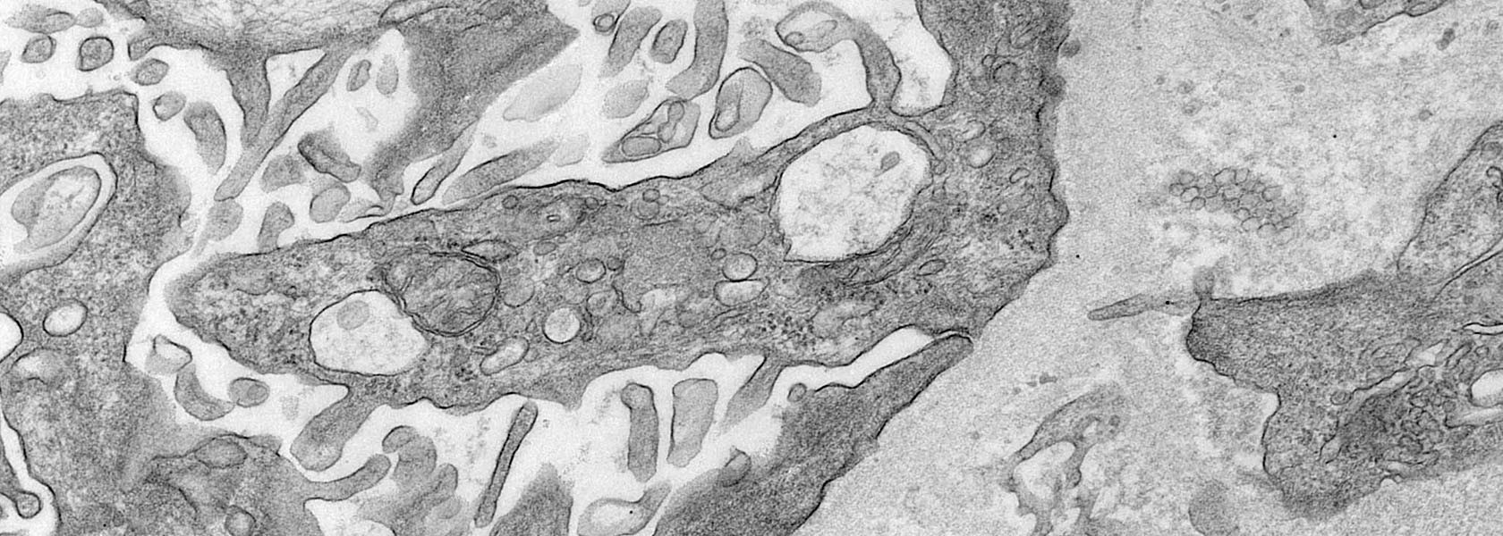

Clinical and Molecular Nephrology
We aim at a better understanding of podocytopathies, the complex principles of kidney diseases,
immune-tissue interactions, and interorgan signaling.

“Enabling and performing excellence in clinical and molecular nephrology.”
Prof. Dr. Tobias B. Huber

“Understanding antibody-mediated kidney disease to pave the way for antigen-specific future treatments.”
Prof. Nicola Tomas
Project details and goals
Targeting immune-mediated kidney diseases
Autoimmune kidney diseases are a common cause of kidney failure and are responsible for its most aggressive and progressive forms, mainly in younger individuals. Autoantibodies play key pathogenic roles in most of these disorders and multiple disease-specific target antigens have been identified – circumstances that have shifted treatment strategies from largely unspecific immunosuppression towards B and plasma cell-targeted treatments.
However, such treatments still involve broad immunosuppression with potentially severe adverse effects. Our vision is the development and experimental implementation of pathogenesis-based, highly specific therapies for immune-mediated kidney diseases. This involves targeting kidney cells using adeno-associated viral vectors as well as antibody- and nanobody-based strategies to eliminate autoreactive B and plasma cells.
Current projects

Podocytopathies
We aim to identify both antibody and non-antibody causative agents in podocytopathies with a minimal change and primary focal-segmental glomerulosclerosis phenotype. In addition, we analyze the tissue response to these factors to identify pathogenic disease mechanisms.

Anti-nephrin autoantibodies
Our teams are currently characterizing the clinical and pathobiological roles of anti-nephrin autoantibodies in podocytopathies. This involves epidemiological studies in large cohorts of patients with different podocytopathies, the biochemical characterization of the autoantibodies and their binding sites within the nephrin molecule, and the establishment of cell-, organoid- and animal-based disease modeling systems.

Gene-Therapy
We are developing novel gene therapy vectors using AAVs to efficiently target kidney glomerular cells, offering a promising new approach to treat antibody-mediated kidney diseases.

Membranous nephropathy (MN)
This project focuses on the conceptualization and preclinical testing of new therapeutic strategies aiming at the removal of pathogenic autoantibodies and autoreactive B-cells.
Publications 2026
Kidney International, Jürgen Floege et al.
Abstract
Treatments that deplete or modulate B cells are in use or being investigated for several immune-mediated glomerular diseases. Kidney Disease: Improving Global Outcomes (KDIGO) convened a Controversies Conference in Panama City, Panama, in June 2025 to review current evidence and identify key gaps in knowledge and research needs to effectively apply such therapies. Availability, effectiveness, and safety of B cell-targeted therapies vary substantially across glomerular diseases. In IgA nephropathy, anti-CD20 therapy (rituximab) has shown limited efficacy, although inhibitors of survival factors BAFF (B cell activating factor) and APRIL (a proliferation-inducing ligand) and anti-CD38 antibodies can lead to reduction in proteinuria and reduction in decline in estimated glomerular filtration rate. In contrast, for membranous nephropathy, anti-CD20 antibodies have become first-line therapy, achieving at least partial remission in most patients by 18 months. In steroid-dependent nephrotic syndrome, rituximab effectively prevents relapses, particularly in children, though benefits are transient. For lupus nephritis, newer approaches including obinutuzumab and chimeric antigen receptor (CAR) T cell therapy have shown promising results, with CAR T cells introducing the possibility of being free of disease activity and treatment for a prolonged time. In antineutrophil cytoplasmic antibody-associated glomerulonephritis, rituximab has proven effective for both induction and maintenance therapy, with ongoing trials investigating CAR T cell approaches. Safety considerations of B cell-targeting therapies vary by intensity of therapy, with conventional anti-CD20 therapy showing favorable safety profiles and CAR T cell therapy requiring careful patient selection because of the potential for cytokine release syndrome and other serious adverse events. Validating biomarkers for patient selection and monitoring is a critical research need, along with optimizing treatment protocols and determining optimal therapy duration.
JCI insight, Ming Huang et al.
Abstract
Although the pathogenic role of autoantibodies targeting the podocyte protein THSD7A in membranous nephropathy (MN) is well described, the consequences of autoantibody binding for podocyte homeostasis and the function of THSD7A remain unclear. Here, we induced an MN model in control and podocyte-specific Thsd7a-KO (Thsd7a-/-) mice using rabbit anti-THSD7A antibodies, followed by transcriptome and proteome analyses. Anti-THSD7A antibodies in WT mice caused significant loss of key slit diaphragm (SD) proteins, such as nephrin and NEPH1, without transcriptional downregulation. Glomeruli showed substantial transcriptomic and proteomic reconfiguration indicative of extensive podocyte injury, including disruptions in podocyte adhesion, cytoskeletal dynamics, and marked upregulation of ubiquitin-proteasome system components, cathepsins, and ADAM proteases. Notably, experiments in C3-deficient mice revealed that proteolytic activation and SD protein loss are driven by complement-independent pathways. Thsd7a-/- mice only displayed a mild phenotype under basal conditions, and they were completely protected from MN development upon anti-THSD7A antibody transfer. Finally, interactome analysis identified a protein complex, including THSD7A and integrin α3, linking THSD7A complexes to pathogenic regulation of cytoskeleton, adhesion, and membrane signaling in MN. Thus, anti-THSD7A antibodies induce profound molecular reconfiguration, including dysregulated proteolytic systems via a complement-independent pathway, revealing potential therapeutic targets in MN.
Kidney Int Rep., Felicitas E. Hengel et al.
Abstract
Introduction: The identification of autoantibodies against nephrin, a key signaling protein of the podocyte slit diaphragm, and their pathogenic role in patients with minimal change disease (MCD) has substantially changed our understanding of primary podocytopathies. However, details on underlying immune pathophysiological mechanisms such as the relation of anti-nephrin autoantibodies with immune cell subsets and therapy response remain to be determined.
Methods: In this prospective study, we evaluated blood samples from adults with newly diagnosed, biopsy-proven MCD for circulating anti-nephrin antibodies using immunoprecipitation (IP) and B-cell subpopulations by fluorescence-activated cell sorting. Samples were collected at diagnosis (t0) and after 8 weeks of steroid therapy (t1).
Results: We detected anti-nephrin antibodies in 12 of 17 (70.6%) therapy-naïve patients with MCD at t0. All 12 patients (100%) positive for anti-nephrin antibodies achieved complete remission with no anti-nephrin antibodies detectable after 8 weeks of steroid treatment (t1). Two of 5 anti-nephrin negative patients (40%) did not reach clinical remission at t1. Anti-nephrin positive patients exhibited a larger fraction of plasmablasts than anti-nephrin negative patients at t0, albeit not statistically significant. Plasmablasts, naïve B cells, and transitional B cells significantly decreased, whereas marginal zone-like B cells increased within 8 weeks of steroid treatment in anti-nephrin positive patients (t1).
Conclusion: Our study shows, for the first time, a therapeutically relevant short-term effect of steroids on the B-cell compartment and anti-nephrin antibody levels, explaining the high clinical response rate in patients with anti-nephrin-associated MCD.
Publications 2025
Kidney Int., Gunther Zahner et al.
Abstract
Introduction: The discovery of anti-phospholipase A2 receptor 1 (PLA2R1) autoantibodies in patients with membranous nephropathy (MN) has led to a paradigm change in diagnosis, monitoring, risk prediction, and therapy of this autoimmune disease. However, there is only limited data on the pathogenicity of these autoantibodies.
Methods: We purified the IgG from sera of patients with PLA2R1-associated MN and injected it into Rag2-deficient mice (mice lacking functional adaptive immunity) expressing human PLA2R1 in their podocytes. The IgG-depleted serum from the same patients and IgG purified from healthy individuals served as controls.
Results: Two weeks after transfer, mice receiving anti-PLA2R1 IgG (largely IgG4), but not IgG-depleted serum or control IgG, developed proteinuria and showed granular deposition of human IgG and complement components of both the classical and alternative pathways in glomeruli by immunofluorescence and podocyte foot process effacement and subepithelial electron dense deposits in electron microscopy. There was no mouse IgG deposited. The IgG eluted from glomeruli of anti-PLA2R1 IgG-injected mice exclusively recognized PLA2R1.
Conclusions: Our findings provide evidence for a direct pathogenic role of anti-PLA2R1 autoantibodies, supporting the development of treatments aiming at the reduction of anti-PLA2R1 autoantibody levels.
Kidney Int Rep., Felicitas E. Hengel et al.
Abstract
Introduction: Circulating autoantibodies against the podocyte surface protein nephrin have recently been described in patients with podocytopathies, that is, minimal change disease, primary focal segmental glomerulosclerosis, and childhood idiopathic nephrotic syndrome. Their high specificity for podocytopathies in combination with a strong correlation with disease activity hold the potential for a non-invasive diagnosis, but prospective data are lacking.
Methods: Here, we describe 3 patients with contraindications or unwillingness for a kidney biopsy, hampering a timely histological diagnosis and choice of appropriate therapy.
Results: In all patients, antinephrin autoantibodies were detected by quantitative immunoprecipitation, prompting the initiation of adequate treatment. These interventions induced a decrease in antinephrin autoantibody levels and clinical remission.
Conclusion: Our study highlights the potential of antinephrin autoantibody measurement for a noninvasive diagnosis of antinephrin-associated podocytopathy.
Nature, Malte Kuehl et al.
Abstract
The expression and location of proteins in tissues represent key determinants of health and disease. Although recent advances in multiplexed imaging have expanded the number of spatially accessible proteins, the integration of biological layers (that is, cell structure, subcellular domains and signalling activity) remains challenging. This is due to limitations in the compositions of antibody panels and image resolution, which together restrict the scope of image analysis. Here we present pathology-oriented multiplexing (PathoPlex), a scalable, quality-controlled and interpretable framework. It combines highly multiplexed imaging at subcellular resolution with a software package to extract and interpret protein co-expression patterns (clusters) across biological layers. PathoPlex was optimized to map more than 140 commercial antibodies at 80 nm per pixel across 95 iterative imaging cycles and provides pragmatic solutions to enable the simultaneous processing of at least 40 archival biopsy specimens. In a proof-of-concept experiment, we identified epithelial JUN activity as a key switch in immune-mediated kidney disease, thereby demonstrating that clusters can capture relevant pathological features. PathoPlex was then used to analyse human diabetic kidney disease. The framework linked patient-level clusters to organ disfunction and identified disease traits with therapeutic potential (that is, calcium-mediated tubular stress). Finally, PathoPlex was used to reveal renal stress-related clusters in individuals with type 2 diabetes without histological kidney disease. Moreover, tissue-based readouts were generated to assess responses to inhibitors of the glucose cotransporter SGLT2. In summary, PathoPlex paves the way towards democratizing multiplexed imaging and establishing integrative image analysis tools in complex tissues to support the development of next-generation pathology atlases.
© 2025. The Author(s).
Kidney Int., Felicitas E. Hengel et al.
Abstract
Introduction: Autoantibodies against the podocyte protein nephrin were recently identified in a pediatric cohort primarily comprising steroid-sensitive (SSNS) and steroid-dependent (SDNS) nephrotic syndrome (NS). However, their prevalence across all NS subtypes, particularly in steroid-resistant nephrotic syndrome (SRNS), and their relation to therapy response need to be determined to advance pathophysiological understanding and refine treatment strategies.
Methods: A multicenter cohort study measuring anti-nephrin autoantibodies in samples from children with SSNS, SDNS, nongenetic and genetic SRNS was conducted.
Results: Sixty-nine of 101 (68%) patients with SSNS, 19 of 67 (28%) patients with SDNS, 14 of 103 patients (14%) with non-genetic SRNS, and 1 of 62 patients (2%) with genetic SRNS were positive for anti-nephrin autoantibodies. The prevalence of anti-nephrin autoantibodies increased with presence of active disease in cases of SSNS and SDNS. Within the group of non-genetic SRNS patients with active disease, anti-nephrin positivity was found in 13 of 74 (18%) patients responding to intensified immunosuppression compared to none of 17 patients with multidrug-resistant SRNS.
Conclusions: The prevalence of anti-nephrin antibodies is substantially higher in children with steroid responsive NS than in those with SRNS, suggesting that anti-nephrin antibodies primarily drive SSNS/SDNS. In contrast, NS due to podocyte gene mutations is primarily genotype-caused. Anti-nephrin autoantibodies may serve as a positive prognostic marker in pediatric NS, indicating a favorable response to immunosuppressive therapy.
Copyright © 2025 International Society of Nephrology. Published by Elsevier Inc. All rights reserved.
J Clin Invest., Felicitas E. Hengel et al.
No abstract available
Team


Prof. Dr. med. Tobias Huber
Head



Prof. Nicola Tomas
Head